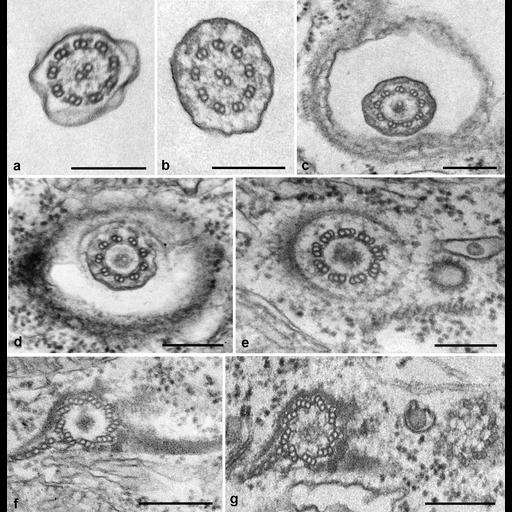
A composite of cross sections through the cilium-basal body complex extending from the cilium to the transition zone between the cilium and basal body and ending at the proximal tip of the basal body. Bars = 0.2µm. a. Cross section of cilium. TEM taken 8/4/65 by R. Allen with RCA EMU3F operating at 50kV. b. Cross section near the proximal end of the cilium. Triangular densities link the doublets to the membrane. TEM taken 7/23/65 by R. Allen with RCA EMU3F operating at 50kV. c. Level of singlet ends and narrowing of plasma membrane around the axoneme. TEM Taken 8/7/65 by R. Allen with RCA EMU3F operating at 50kV. d. Similar to c showing the curving of the plasma membrane and the enclosed alveoli. TEM taken 8/7/65 by R. Allen with RCA EMU3F operating at 50kV. e. Distal end of basal body showing 9 triplets and the electron opaque central core. The coated end of a parasomal sac lies anterior to the basal body. TEM taken 8/9/65 by R. Allen with RCA EMU3F operating at 50kV. f. Toward the proximal end of the basal body. TEM taken on 8/6/65 by R. Allen with RCA TEM. g. Proximal ends of two basal bodies showing the cartwheel end in one and just distal to the cartwheel in the second basal body. kd, kinetodesmal fiber; pc mt, postciliary microtubules. TEM taken 8/7/65 by R. Allen with RCA TEM. Published in J. Cell Biol. 40:716-733, 1969. Adapted with permission. The negative was printed to paper and the image was scanned to Photoshop. This digitized image is available for qualitative analysis. Additional information available at (http://www5.pbrc.hawaii.edu/allen/).
Standard glutaraldehyde fixation followed by osmium tetroxide, dehydrated in alcohol and embedded in an epoxy resin. Microtome sections prepared at approximately 75nm thickness. Additional information available at (http://www5.pbrc.hawaii.edu/allen/).
| Spatial Axis | Image Size | Pixel Size |
|---|---|---|
| X | 2108px | —— |
| Y | 2168px | —— |