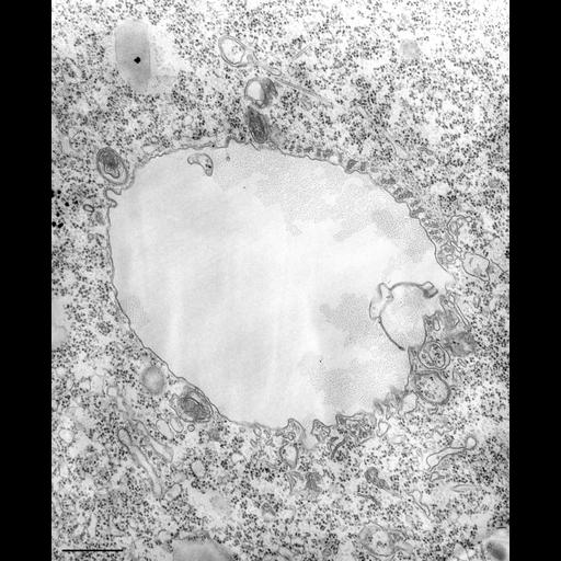
The nascent digestive vacuole forms as the membrane of flattened vesicles fuse with the single membrane of the cytopharynx between the lamellae (2 microtubules forming a lamella) that are connected along their length to the membrane at the cytopharynx. Another portion of the forming vacuole is associated with the ends of the 4 element- (4 mt) and 2 element- (2 mt) microtubular ribbons that come from the oral ribs. These ribbons are covered with specialized cytoplasm and do not take part in moving discoidal vesicles to the vesicle fusion region. One feature of the nascent vacuole in Tetrahymena that has not been observed in Paramecium is the presence of coated pits extending from the vacuole membrane. Finally, vesicles previously called lysosomes, which to me resemble the late endosomes, acidosomes, of Paramecium, dock at the growing vacuole. For a review of the literature of phagocytosis in Tetrahymena see Nilsson, Biochemistry and Physiology of Protozoa, 2nd ed., Academic Press, pp. 339-379, 1979; Sattler and Staehelin, J. Ultrastructure Res. 66:132-150, 1979; and Baumert et al., Eur. J. Protistol. 34:291-300, 1998. TEM taken on 8/15/67 by R. Allen with Philips 200 operating at 60kV. Neg 19,200X. Bar = 0.5µm. The negative was printed to paper and the image was scanned to Photoshop. This digitized image is available for qualitative analysis. There is a high resolution version of this image in the library (CIL:39720) which is available for quantitative analysis. Additional information available at (http://www5.pbrc.hawaii.edu/allen/).
Standard glutaraldehyde fixation followed by osmium tetroxide, dehydrated in alcohol and embedded in an epoxy resin. Microtome sections prepared at approximately 75nm thickness. Additional information available at (http://www5.pbrc.hawaii.edu/allen/).
| Spatial Axis | Image Size | Pixel Size |
|---|---|---|
| X | 3069px | —— |
| Y | 3760px | —— |