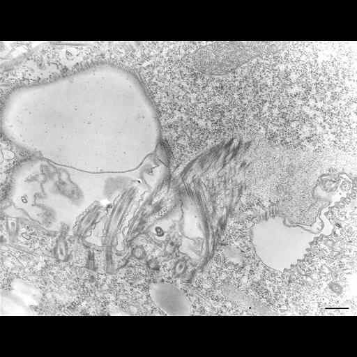
Some accessory microtubules from the proximal ends of membranelle basal bodies extend upward and curve over the left wall of the buccal cavity as can be followed in this micrograph. Some fibrous components (the posterior connectives, see Smith and Buhse, Trans. Am. Microsc. Soc. 102:264-271, 1983) from the membranelles pass into the specialized cytoplasm where it forms an electron opaque mass. The bottom of the buccal cavity bordered by oral ribs opens through the cytosome into the nascent digestive vacuole. The cytopharynx is the site of lamellar microtubules (1 or 2 microtubules per lamella) which direct flattened vesicles to the growing vacuole’s fusion site. TEM taken on 8/16/67 by R. Allen with Philips 200 operating at 60kV. Neg. 12,400X. Bar = 0.5µm. The negative was printed to paper and the image was scanned to Photoshop. This digitized image is available for qualitative analysis. There is a high resolution version of this image in the library (CIL:39717) which is available for quantitative analysis. Additional information available at (http://www5.pbrc.hawaii.edu/allen/).
Standard glutaraldehyde fixation followed by osmium tetroxide, dehydrated in alcohol and embedded in an epoxy resin. Microtome sections prepared at approximately 75nm thickness. Additional information available at (http://www5.pbrc.hawaii.edu/allen/).
| Spatial Axis | Image Size | Pixel Size |
|---|---|---|
| X | 3968px | —— |
| Y | 3056px | —— |