Licensing

Attribution_by_nc_nd
Attribution Non-Commercial; No Derivatives:This image is licensed under a Creative Commons Attribution, Non-Commercial, No Derivatives License. View License Deed | View Legal Code
Comments: Powered by Pnyxe

Description

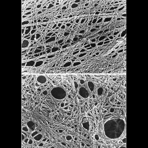
Figures 440 (upper) and 441 (lower) from Chapter 16 (Cytoplasmic matrix and cytoskeleton) of 'The Cell, 2nd Ed.' by Don W. Fawcett M.D. Examples of the dense cytoskeletal network in the periphery of cells in vitro. In the upper panel, two microtubules traverse the meshwork of actin filaments. This cell was prepared by treated with Triton to extract soluble proteinaceous material, washed, quick frozen with liquid helium, deep etched, and rotary shadowed. Image by John Heuser. A PDF copy of the accompanying chapter is available on the ASCB's BioEDUCATE website.

Biological Sources

NCBI Organism Classification
Mus musculus
(house mouse)
Cell Type
fibroblast
Cell Line
3T3-Swiss albino
Cellular Component
actin cytoskeleton
cytoplasmic microtubule

Attribution

Names
Don W. Fawcett
John Heuser
Published
ISBN 0721635849
Link
http://www.ascb.org/bioeducate/Fawcett...

Grouping

This image is part of a group.
Data Qualifiers
processed data

Sample Preparation

Methods
detergent permeabilized
FBbi:00000621
Relation To Intact Cell
dispersed cells in vitro

Dimensions

Spatial Axis Image Size Pixel Size
X 891px ——
Y 1257px ——
*CIL – Cell Image Library accession number. Please use this to reference an image.