Alternate header for print version


Licensing
Attribution Non-Commercial; No Derivatives:This image is licensed under a Creative Commons Attribution, Non-Commercial, No Derivatives License. View License Deed | View Legal Code
tweet  
*CIL – Cell Image Library accession number. Please use this to reference an image.

CIL:36067*  Cite 
Description

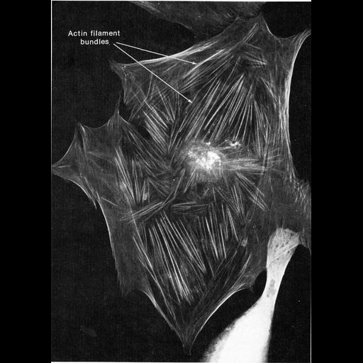
A thinly-spread human skin fibroblast stained by indirect immunofluorescence with actin-specfic antibody. Actin bundles correspond with stress fibers, extending out into the periphery of the cell. Figure 435 from Chapter 16 (Cytoplasmic matrix and cytoskeleton) of 'The Cell, 2nd Ed.' by Don W. Fawcett M.D. Image from Elias Lazarides, J. Cell Biol. 65:549-61, 1975, PMID: 1094020. A PDF copy of the accompanying chapter is available on the ASCB’s BioEDUCATE website.

Biological Sources
NCBI Organism Classification
Homo sapiens
Cell Type
fibroblast
Cellular Component
actin filament bundle
stress fiber
Biological Context
Biological Process
actin cytoskeleton organization
Attribution
Names
Don W. Fawcett
Elias Lazarides
Published
ISBN 0721635849
Link
http://www.ascb.org/bioeducate/FawcettTheCell...
Citation
Digital Object Identifier (DOI)
doi:10.7295/W9CIL36067
Archival Resource Key (ARK)
ark:/b7295/w9cil36067
Grouping This image is part of a group.
Imaging
Image Type
recorded image
Image Mode
fluorescence microscopy
Parameters Imaged
fluorescence emission
Source of Contrast
distribution of epitope
Visualization Methods
primary antibody plus labeled secondary antibody
Data Qualifiers
processed data
Sample Preparation
Methods
crosslinking-fixative fixed tissue
detergent permeabilized
Relation To Intact Cell
dispersed cells in vitro
Dimensions
Spatial Axis Image Size Pixel Size
X 888px ——
Y 1240px ——