Licensing
Attribution Non-Commercial; No Derivatives:This image is licensed under a Creative Commons Attribution, Non-Commercial, No Derivatives License. View License Deed | View Legal Code
Comments:
Powered by Pnyxe
Description
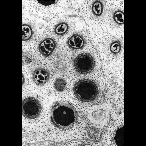
Figure 421 from Chapter 16 (Cytoplasmic matrix and cytoskeleton) of 'The Cell, 2nd Ed.' by Don W. Fawcett M.D. Horizontal section through the apical cytoplasm of a Sertoli cell in the testis of the toad Buro marinus. Microtubules in these polarized cells are abundant and consistent in their orientation. The Sertoli cells serve as nurse cells for maturing spermatozoa, and late in sperm maturation, the sperm heads occupy deep recesses in the apical part of the Sertoli cells. A number of sperm at different levels are apparent in this section, surrounded by the cytoplasm of the Sertoli cell. A PDF copy of the accompanying chapter is available on the ASCB's BioEDUCATE website.
Biological Sources
- NCBI Organism Classification
- Bufo marinus
- (Cane Toad)
- Cell Type
- Sertoli cell
- sperm
- Cellular Component
- microtubule
- apical part of cell
Biological Context
Attribution
- Name
- Don W. Fawcett
- Published
- ISBN 0721635849
- Link
- http://www.ascb.org/bioeducate/Fawcett...
Grouping
This image is part of a group.Imaging
- Image Type
- transmission electron microscopy (TEM)
- Imaging Mode
- illumination by electrons
- detection of electrons
- Parameters Imaged
- electron density
- Source of Contrast
- stain with broad specificity
- Visualization Methods
- electron-dense stain
- Data Qualifiers
- processed data
Sample Preparation
- Methods
- crosslinking-fixative fixed tissue
- tissue in plastic embedment
- Relation To Intact Cell
- microtome-sectioned tissue
Dimensions
| Spatial Axis | Image Size | Pixel Size |
|---|---|---|
| X | 879px | —— |
| Y | 1251px | —— |
*CIL – Cell Image Library accession number. Please use this to reference an image.