Alternate header for print version


Licensing
This image is copyright protected. Any public or private use of this image is subject to prevailing copyright laws. Please contact the content provider of this image for permission requests.
tweet  
*CIL – Cell Image Library accession number. Please use this to reference an image.

CIL:36037*  Cite 
Description

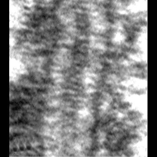
Transmission electron micrograph showing the characteristic features of the synaptonemal complex in a thin section of a Drosophila oocyte.

Technical Details

Ovaries were fixed in glutaraldehye, post-fixed in Osmium tetroxide, embedded in plastic, and thin sections stained with uranyl acetate and lead citrate. Thin sections were examined with an AEI 801 electron microscope.

Biological Sources
NCBI Organism Classification
Drosophila melanogaster
Cell Type
oocyte
Cellular Component
synaptonemal complex
Biological Context
Biological Process
female meiosis
Attribution
Names
Lorinda Anderson
Suzanne Royer
Scott Page
Kim McKim
Ann Lai
Mary Lilly
R Scott Hawley
Published
LK Anderson et al. (2005) Proc Natl Acad Sci 102:4482-4487
Pubmed
15767569
Citation
Digital Object Identifier (DOI)
doi:10.7295/W9CIL36037
Archival Resource Key (ARK)
ark:/b7295/w9cil36037
Imaging
Image Type
recorded image
Image Mode
transmission electron microscopy (TEM)
Parameters Imaged
electron density
Source of Contrast
differences in adsorption or binding of stain
Visualization Methods
osmium tetroxide
uranyl salt
lead salt
Data Qualifiers
processed data
Sample Preparation
Methods
glutaraldehyde fixed tissue
tissue in plastic embedment
Relation To Intact Cell
microtome-sectioned tissue
Dimensions
Spatial Axis Image Size Pixel Size
X 900px ——
Y 1020px ——