Alternate header for print version


Licensing
This image is copyright protected. Any public or private use of this image is subject to prevailing copyright laws. Please contact the content provider of this image for permission requests.
tweet  
*CIL – Cell Image Library accession number. Please use this to reference an image.

CIL:36025*  Cite 
Description

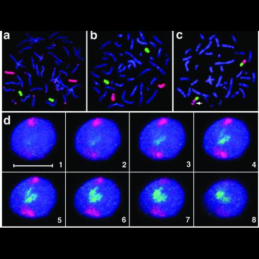
Fluorescence in situ hybridization (FISH) of chromosome spreads and interphase nuclei of lymphoblastoid cell of higher primates using probes specific to individual chromosomes. The figure illustrates the concept of chromosome territories with individual chromosomes occupying distinct spatial territories during interphase. Top row identifies chromosomes 16 (red) and 20 (green) in gorilla (a) and human (b). d shows selected z-slices of a marmoset interphase nucleus revealing the territories occupied by chromosome 18(red) and 19 (green). DNA is shown in blue.

Technical Details

Epstein-Barr virus-transformed lymphoblastoid cells were fixed in paraformaldehyde, processed for FISH and z-series recorded with a Zeiss LSM 410 confocal microscope.

Biological Sources
NCBI Organism Classification
Gorilla
Homo sapiens
Callithrix jacchus
Cell Type
lymphoblastoid
Cellular Component
nuclear chromosome
Attribution
Names
Hideyuki Tanabe
Stefan Muller
Michaela Neusser
Johann von Hase
Enzo Calcagno
Marion Cremer
Irina Solovei
Christoph Cremer
Thomas Cremer
Published
H. Tanabe et al. 2002 Proc Natl Acad Sci 99:4424-4429
Pubmed
11930003
Link
chromosome territory
Citation
Digital Object Identifier (DOI)
doi:10.7295/W9CIL36025
Archival Resource Key (ARK)
ark:/b7295/w9cil36025
Imaging
Image Type
recorded image
Image Mode
fluorescence microscopy
confocal microscopy
Parameters Imaged
fluorescence emission
Source of Contrast
distribution of a specific nucleic acid sequence
Processing History
montage
Data Qualifiers
processed data
Sample Preparation
Methods
formaldehyde fixed tissue
Dimensions
Spatial Axis Image Size Pixel Size
X 519px ——
Y 395px ——