Alternate header for print version


Licensing
Attribution Non-Commercial; No Derivatives:This image is licensed under a Creative Commons Attribution, Non-Commercial, No Derivatives License. View License Deed | View Legal Code
tweet  
*CIL – Cell Image Library accession number. Please use this to reference an image.

CIL:35980*  Cite 
Description

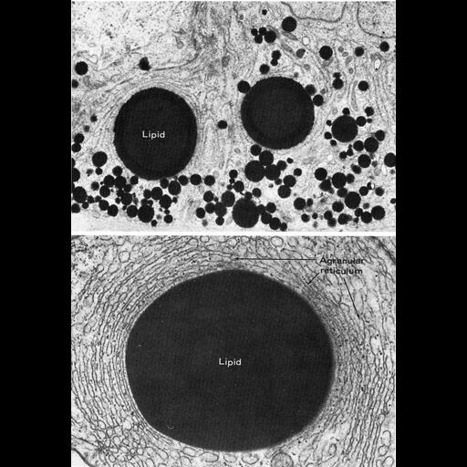
Figures 358 (upper) and 359 (lower) from Chapter 15 (Cytoplasmic Inclusions) of 'The Cell, 2nd Ed.' by Don W. Fawcett M.D. In some species, Sertoli cells, lipid droplets are abundant. Upper panel: multiple small and medium-sized lipid droplets at the base of Sertoli cells from the gerenuk (Litocranius walleri). Lower panel: Lipid droplet in a Sertoli cell from the ram (Ovis aries), surrounded by smooth reticulum. Figures from Fawcett in Handbook of physiology, Sect. 7, Vol. 5, AM. Phyiol. Soc., Washington D.C., 1975. A PDF copy of the accompanying chapter is available on the ASCB’s BioEDUCATE website.

Biological Sources
NCBI Organism Classification
Litocranius walleri
Ovis aries
Cell Type
Sertoli cell
Cellular Component
lipid particle
smooth endoplasmic reticulum
Biological Context
Biological Process
lipid storage
Attribution
Names
Don W. Fawcett
Published
ISBN 0721635849
Link
http://www.ascb.org/bioeducate/FawcettTheCell...
Citation
Digital Object Identifier (DOI)
doi:10.7295/W9CIL35980
Archival Resource Key (ARK)
ark:/b7295/w9cil35980
Grouping This image is part of a group.
Imaging
Image Type
transmission electron microscopy (TEM)
Image Mode
illumination by electrons
detection of electrons
Parameters Imaged
electron density
Source of Contrast
stain with broad specificity
Visualization Methods
osmium tetroxide
Data Qualifiers
processed data
Dimensions
Spatial Axis Image Size Pixel Size
X 894px ——
Y 1285px ——