Alternate header for print version


Licensing
Attribution Non-Commercial; No Derivatives:This image is licensed under a Creative Commons Attribution, Non-Commercial, No Derivatives License. View License Deed | View Legal Code
tweet  
*CIL – Cell Image Library accession number. Please use this to reference an image.

CIL:35970*  Cite 
Description

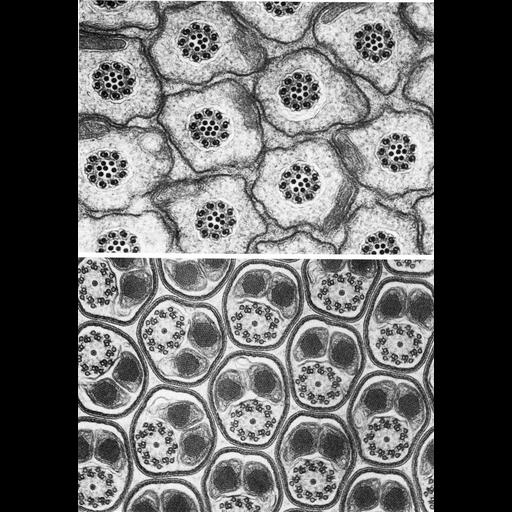
Figures 342 (upper) and 343 (lower) from Chapter 14 (Sperm Flagellum) of 'The Cell, 2nd Ed.' by Don W. Fawcett M.D. Cross sections of spermatozoa from the caddis fly (Polycentropus, upper) and mosquito (Culex, lower) provide examples of variation in the microtuble organization of motile flagella. In contrast to common 9 + 2, or 9 + 9 + 2, the caddis fly shows a 9 + 7 organization, whereas the mosquito exhibits 9 + 9 + 1 pattern. Images by David Phillips; Fig 343 from Phillips, J. Cell Biol. 40:28-43, 1969. A PDF copy of the accompanying chapter is available on the ASCB’s BioEDUCATE website.

Biological Sources
NCBI Organism Classification
Polycentropus
Culex
Cell Type
sperm
Cellular Component
axoneme
microtubule-based flagellum
Biological Context
Biological Process
fertilization
Attribution
Names
Don W. Fawcett
David Phillips
Published
ISBN 0721635849
Link
http://www.ascb.org/bioeducate/FawcettTheCell...
Citation
Digital Object Identifier (DOI)
doi:10.7295/W9CIL35970
Archival Resource Key (ARK)
ark:/b7295/w9cil35970
Grouping This image is part of a group.
Imaging
Image Type
transmission electron microscopy (TEM)
Image Mode
illumination by electrons
detection of electrons
Parameters Imaged
electron density
Source of Contrast
stain with broad specificity
Visualization Methods
osmium tetroxide
Data Qualifiers
processed data
Dimensions
Spatial Axis Image Size Pixel Size
X 880px ——
Y 1266px ——