Alternate header for print version


Licensing
Public Domain: This image is in the public domain and thus free of any copyright restrictions. However, as is the norm in scientific publishing and as a matter of courtesy, any user should credit the content provider for any public or private use of this image whenever possible. Learn more
tweet  
*CIL – Cell Image Library accession number. Please use this to reference an image.

CIL:35856*  Cite 
Description

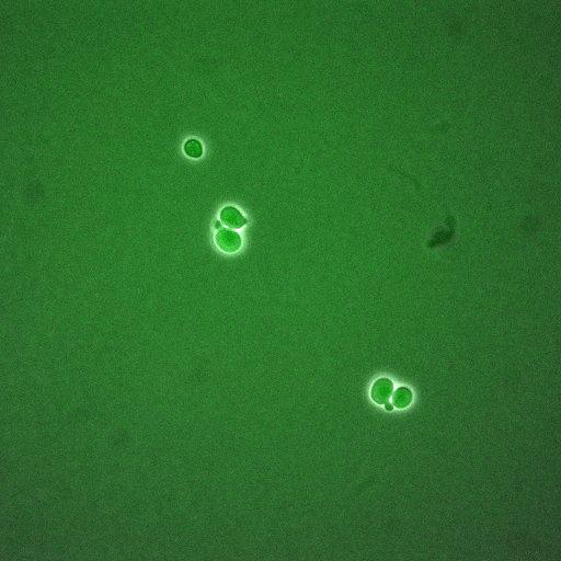
A time lapse experiment of Saccharomyces cerevisiae expressing GFP tagged Cdc15, a protein kinase involves in cytokinesis. These phase and GFPimages are part of an image group containing CIL 35840-35859. Note that there are additional groups of images of other cell cycle regulator proteins by the same authors available in the Library.

Technical Details

The images were acquired using a 60x 1.42 NA objective lens on Delta Vision system equipped with CoolSNAP_HQ2 / HQ2-ICX285. Please read the microscopy section of the referenced paper for details before image analysis.

Biological Sources
NCBI Organism Classification
Saccharomyces cerevisiae
Cellular Component
CDC15 Protein
cellular bud neck
spindle pole body
Attribution
Names
David Ball
Jean Peccoud
Published
PLoS One. 2011;6(10):e26272. Epub 2011 Oct 26.
Pubmed
22046265
Citation
Digital Object Identifier (DOI)
doi:10.7295/W9CIL35856
Archival Resource Key (ARK)
ark:/b7295/w9cil35856
Grouping This image is part of a group.
Imaging
Image Type
recorded image
Image Mode
fluorescence microscopy
phase contrast microscopy
Parameters Imaged
fluorescence emission
elastic scattering of photons
Source of Contrast
distribution of a specific protein
differences in amount of elastic light scattering
Visualization Methods
EGFP
visualization of contiguous regions
Data Qualifiers
raw, unprocessed data
suitable for spatial measurements
Sample Preparation
Methods
living tissue
Relation To Intact Cell
dispersed cells in vitro
Dimensions
Spatial Axis Image Size Pixel Size
X 512px 213.4nm
Y 512px 213.4nm
Time 300 seconds 61