Licensing
This image is copyright protected. Any public or private use of this image is subject to prevailing copyright laws. Please contact the content provider of this image for permission requests.
Comments:
Powered by Pnyxe
Description
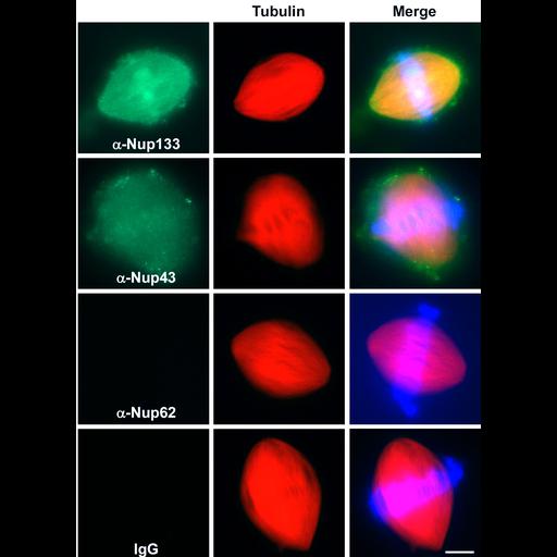
Distribution of Nup133, Nup43 and Nup62 (green) and tubulin (blue) in spindles assembled in vitro from Xenopus oocyte extracts. Chromosomes are stained with DAPI (blue). See Orjalo et al 2006. Mol Biol Cell 17:3806-3808.
Technical Details
Xenopus egg extracts were mixed with sperm chromatin, treated to induce spindle formation, antibodies added, and the preparations fixed with paraformaldehyde, and centrifuged onto cover slips through a glycerol cushion, and processed for immunofluorescence. z-stacks of images were recorded using a Zeiss Axioskop microscope with 63x !.4 NA objective lens. See Fig 3A in Orjalo et al. 2006 Mol Biol Cell 17:3806-3818.
Biological Sources
- NCBI Organism Classification
- Xenopus laevis laevis
- Cell Type
- oocyte
- Cellular Component
- spindle
Biological Context
- Biological Process
- mitosis
Attribution
- Names
- Arturo Orjalo
- Alexei Arnaoutov
- Zhouxin Shen
- Yekaterina Boyarchuk
- Samantha Zeitlin
- Beatriz Fontoura
- Steven Briggs
- Mary Dasso
- Douglass Forbes
- Published
- Orjalo et al. 2006 The Nup107-160 nucleoporin complex is required for correct bipolar spindle assembly. Mol Biol Cell 17:3806-3818
- Pubmed
- 16807356
Grouping
This image is part of a group.Imaging
- Image Type
- recorded image
- Imaging Mode
- fluorescence microscopy
- Parameters Imaged
- fluorescence emission
- Source of Contrast
- distribution of epitope
- differences in adsorption or binding of stain
- Visualization Methods
- 4',6-diamidino-2-phenylindole (DAPI)
- Tetramethyl rhodamine (TRITC)
- Processing History
- montage
- Data Qualifiers
- processed data
Sample Preparation
- Methods
- coagulating-fixative fixed tissue
- formaldehyde fixed tissue
- Relation To Intact Cell
- isolated subcellular component
Dimensions
| Spatial Axis | Image Size | Pixel Size |
|---|---|---|
| X | 1582px | —— |
| Y | 2178px | —— |
*CIL – Cell Image Library accession number. Please use this to reference an image.