Licensing

Pd
Public Domain: This image is in the public domain and thus free of any copyright restrictions. However, as is the norm in scientific publishing and as a matter of courtesy, any user should credit the content provider for any public or private use of this image whenever possible. Learn more
Comments: Powered by Pnyxe

Description

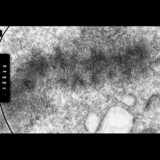
Metaphase-arrested CHO cells were swollen in 75 mM KCL, fixed with formaldehyde, post-fixed with OsO4, embedded, and 0.25 micron sections examined by High Voltage EM. This image was recorded with a specimen tilt of 45 degrees. Grouped with it is one recorded at 55 degrees, providing an oblique stereo view of chromosome organization.

Technical Details

Colcemid-arrested cells were swollen in hypotonic 75mM KCl, fixed with 4% HCOH, post-fixed with OsO4, embedded in plastic, and 'thick' 0.25 micron sections prepared. Sections were recorded using the Wisconsin HVEM at 1 MeV. See Ris (1978) Preparation of chromatin and chromosomes for electron microscopy. Meth Cell Biol 18:229-246; Ris (1981) Stereoscopic electron microscopy of chromosomes. Meth Cell Biol 22:77-96.

Biological Sources

NCBI Organism Classification
Cricetulus griseus
Cell Type
ovary
Cell Line
CHO
Cellular Component
nuclear chromosome

Biological Context

Biological Process
mitosis

Attribution

Name
Hans Ris

Grouping

This image is part of a group.

Imaging

Image Type
film
Imaging Mode
FBbi:00000622
Parameters Imaged
electron density
Source of Contrast
differences in adsorption or binding of stain
Visualization Methods
osmium tetroxide
Processing History
unprocessed raw data

Sample Preparation

Methods
formaldehyde fixed tissue
tissue in plastic embedment
Relation To Intact Cell
microtome-sectioned tissue

Dimensions

Spatial Axis Image Size Pixel Size
X 4144px 1.0nm
Y 2781px 1.0nm
*CIL – Cell Image Library accession number. Please use this to reference an image.