Alternate header for print version


Licensing
Public Domain: This image is in the public domain and thus free of any copyright restrictions. However, as is the norm in scientific publishing and as a matter of courtesy, any user should credit the content provider for any public or private use of this image whenever possible. Learn more
tweet  
*CIL – Cell Image Library accession number. Please use this to reference an image.

CIL:35529*  Cite 
Description

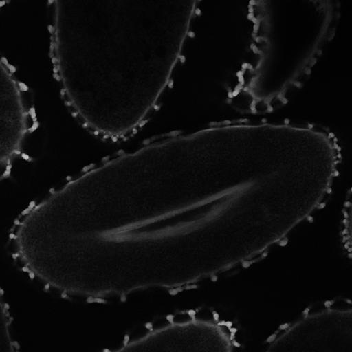
Lilium longiflorum (Easter Lily) pollen. This is a thin optical section through the center of the desiccated stage of the mature pollen showing autofluorescence or harmonic generation of intrinsic structures. The brightest signal is from the outer layer, the exine and raised pilae.

Technical Details

Illumination provided by a SpectraPhysics-Newport MaiTai laser with DeepSee dispersion compensation providing pulses of approximately 100 fs duration at 80 MHz. Wavelength set at 760 nm with emission at 465 to 50 nm. Zeiss 710 multi-photon microscope with 63X N.A. 1.4 oil immersion objective. Pollen was removed from the flower, put in a coverslip bottom chamber, covered with tissue culture grade mineral oil, and imaged within 12 hours.

Biological Sources
NCBI Organism Classification
Lilium longiflorum
Cell Type
gamete
Cellular Component
pollen wall
exine
intine
sexine
endexine
Biological Context
Biological Process
pollination
Plant Growth
L mature pollen stage
Attribution
Names
Michael Cammer
Citation
Digital Object Identifier (DOI)
doi:10.7295/W9CIL35529
Archival Resource Key (ARK)
ark:/b7295/w9cil35529
Grouping This image is part of a group.
Imaging
Image Type
recorded image
Image Mode
multi-photon microscopy
Parameters Imaged
fluorescence emission
Source of Contrast
intrinsic mass distribution
Visualization Methods
visualization of contiguous regions
Processing History
unprocessed raw data
Data Qualifiers
raw, unprocessed data
suitable for spatial measurements
Sample Preparation
Methods
unfixed tissue
Relation To Intact Cell
whole mounted tissue