Alternate header for print version


Licensing
Public Domain: This image is in the public domain and thus free of any copyright restrictions. However, as is the norm in scientific publishing and as a matter of courtesy, any user should credit the content provider for any public or private use of this image whenever possible. Learn more
tweet  
*CIL – Cell Image Library accession number. Please use this to reference an image.

CIL:35482*  Cite 
Description

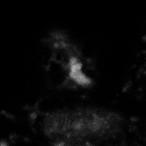
Tripolar mitosis in a liver cancer cell line. Normally, dividing cells form a single metaphase plate of paired chromosomes which are pulled apart by opposing microtubule spindles. The Z series, obtained with a laser scanning confocal microscop,e shows abnormal division by mis-segregated chromosomes. This image group includes other z-series and a 3D surface representation

Technical Details

HuH-7 cells are an immortalized cultured cell line derived from hepatic carcinoma. Cells were grown on 1.5 glass coverslips, fixed with formaldehyde and glutareldehyde, extracted, and labeled for fluorescence microscopy with commercial antibodies to alpha- or beta-tubulin and propidium iodide. The coverslip was mounted on a slide in PBS and glycerol with n-propyl gallate as an antifading agent. Imaging was performed with a BioRad MRC 600 laser scanning confocal microscope with a Kr/Ar laser with lines at 488 and 568 nm. The microscope was a fixed tube length Nikon Diaphot with 60X N.A. 1.4 phase 3 optics. Z series was collected at 0.2 um step sizes. The original XY spatial scale has been lost.

Biological Sources
NCBI Organism Classification
Homo sapiens
Cell Type
hepatocyte
Cell Line
HuH-7
Cellular Component
chromosome
Biological Context
Biological Process
mitotic metaphase
cytokinesis
chromosome segregation
chromosome organization
Human Development Anatomy
liver
Human Disease
sclerosing hepatic carcinoma
Attribution
Names
Michael Cammer
Phyllis Novikoff
Citation
Digital Object Identifier (DOI)
doi:10.7295/W9CIL35482
Archival Resource Key (ARK)
ark:/b7295/w9cil35482
Grouping This image is part of a group.
Imaging
Image Type
recorded image
Image Mode
single-spot confocal microscopy
Parameters Imaged
fluorescence emission
Source of Contrast
distribution of nucleic acids
Visualization Methods
propidium iodide
Processing History
unprocessed raw data
Data Qualifiers
processed data
Sample Preparation
Methods
glutaraldehyde fixed tissue
formaldehyde fixed tissue
Relation To Intact Cell
dispersed cells in vitro
Dimensions
Spatial Axis Image Size Pixel Size
X 512px ——
Y 512px ——
Z 83px 0.2µm