Alternate header for print version


Licensing
Public Domain: This image is in the public domain and thus free of any copyright restrictions. However, as is the norm in scientific publishing and as a matter of courtesy, any user should credit the content provider for any public or private use of this image whenever possible. Learn more
tweet  
*CIL – Cell Image Library accession number. Please use this to reference an image.

CIL:35370*  Cite 
Description

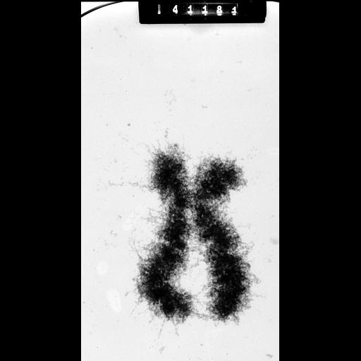
Whole human female metaphase chromosome from metaphase arrested cell swollen in hypotonic medium and imaged at 1 MeV. This image was taken with a specimen tilt of 45 degrees. Grouped with it is an image of the same area at 55 degrees tilt, providing an oblique stereo view of the chromosome.

Technical Details

HeLa cells were metaphase-arrested by a 3hr treatment with colcemid, the cells mechanically broken by passage through a 22-gauge needle and exposed to hypotonic 75 mM KCl for 10 min. Samples were fixed with 3:1 methanol:acetic acid, squashed in 45% acetic acid, transferred to a formvar coated grid, critical point dried, and imaged with the Wisconsin HVEM at 1 MeV. See Ris (1978) Preparation of chromatin and chromosomes for electron microscopy. Meth Cell Biol 18:229-246; Ris (1981) Stereoscopic microscopy of chromosomes. Meth Cell Biol 22:77-96.

Biological Sources
NCBI Organism Classification
Homo sapiens
Cell Type
cevical carcinoma
Cell Line
HeLa
Cellular Component
condensed chromosome
Biological Context
Biological Process
nuclear division
chromosome organization
mitosis
Attribution
Names
Hans Ris, University of Wisconsin, Madison, WI
Citation
Digital Object Identifier (DOI)
doi:10.7295/W9CIL35370
Archival Resource Key (ARK)
ark:/b7295/w9cil35370
Grouping This image is part of a group.
Imaging
Image Type
film
Image Mode
Parameters Imaged
electron density
Source of Contrast
differences in adsorption or binding of stain
Visualization Methods
visualization of contiguous regions
Processing History
unprocessed raw data
Sample Preparation
Methods
coagulating-fixative fixed tissue
critical_point dried specimen
Relation To Intact Cell
whole mounted tissue
Dimensions
Spatial Axis Image Size Pixel Size
X 2610px 1.5µm
Y 4750px 1.5µm