Alternate header for print version


Licensing
Public Domain: This image is in the public domain and thus free of any copyright restrictions. However, as is the norm in scientific publishing and as a matter of courtesy, any user should credit the content provider for any public or private use of this image whenever possible. Learn more
tweet  
*CIL – Cell Image Library accession number. Please use this to reference an image.

CIL:35287*  Cite 
Description

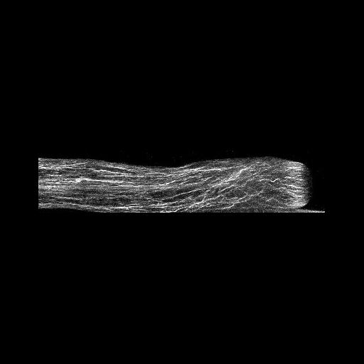
Fixed Lilium pollen tubes were treated with fluorescent phalloidin to label filamentous actin, and confocal images obtained. A strongly labeled actin fringe near the growing tip is present. Other images in the group reveal the distributions of additional cytoskeletal elements and membranous components.

Technical Details

Germinated lily pollen was fixed with paraformaldehyde and glutaraldehye, treated with Alexa-543 phalloidin to label actin, and imaged with a Zeiss 510 meta confocal microscope using a 63x 1.4 NA objective lens. Image is a projection of several confocal x-y slices. See Lovy-Wheeler et al. 2005, Planta 221:95-104.

Biological Sources
NCBI Organism Classification
Lilium formosanum
Cell Type
pollen tube
Cellular Component
actin filament
Biological Context
Biological Process
pollen tube growth
Attribution
Names
Alenka Lovy-Wheeler
Kathleen L Wilsen
Tobias I Baskin
Peter K Hepler
Citation
Digital Object Identifier (DOI)
doi:10.7295/W9CIL35287
Archival Resource Key (ARK)
ark:/b7295/w9cil35287
Grouping This image is part of a group.
Imaging
Image Type
photomultiplier tube (PMT)
Image Mode
confocal microscopy
Parameters Imaged
fluorescence emission
Source of Contrast
differences in adsorption or binding of stain
Visualization Methods
alexa fluor 543
Processing History
projection of slices
Data Qualifiers
processed data
suitable for spatial measurements
Sample Preparation
Methods
formaldehyde fixed tissue
glutaraldehyde fixed tissue
Relation To Intact Cell
whole mounted tissue
Dimensions
Spatial Axis Image Size Pixel Size
X 403px ——
Y 87px ——