Alternate header for print version


Licensing
Public Domain: This image is in the public domain and thus free of any copyright restrictions. However, as is the norm in scientific publishing and as a matter of courtesy, any user should credit the content provider for any public or private use of this image whenever possible. Learn more
tweet  
*CIL – Cell Image Library accession number. Please use this to reference an image.

CIL:35166*  Cite 
Description

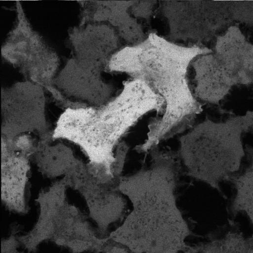
Constitutive expression of GFP (Green Fluorescent protein) in MTLn3 cells, a metastatic rat breast carcinoma line. This time lapse sequence of a single optical section near the substrate shows the cells jostling and the intrinsic movement of organelles that appear dark because they do not contain the fluorescent protein. This cell line is useful both as a cell culture system and also as a source of fluorescent cells that may be used to study metastasis in live rats or mice.

Technical Details

BioRad Radiance 2000 laser scanning confocal microscope with Kr/Ar laser for excitation of permanently transfected GFP at 488 nm. The pixel size is approximate.

Biological Sources
NCBI Organism Classification
Rattus rattus
Cell Type
permanent cell line cell
Cell Line
MTLn3
Cellular Component
cytosol
Biological Context
Biological Process
interphase
Attribution
Names
Maryse Bailly
Michael Cammer
Citation
Digital Object Identifier (DOI)
doi:10.7295/W9CIL35166
Archival Resource Key (ARK)
ark:/b7295/w9cil35166
Imaging
Image Type
single-spot confocal microscopy
Image Mode
fluorescence microscopy
Parameters Imaged
fluorescence emission
Source of Contrast
distribution of a specific protein
Visualization Methods
EGFP
Processing History
unprocessed raw data
Data Qualifiers
raw, unprocessed data
suitable for spatial measurements
Sample Preparation
Methods
living tissue
Relation To Intact Cell
dispersed cells in vitro
Dimensions
Spatial Axis Image Size Pixel Size
X 512px 0.16µm
Y 512px 0.16µm
Time 20 seconds 40