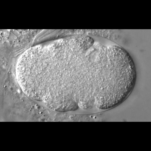
This time lapse series illustrates the early division up to the four-cell stage of a C. elegans embryo depleted of cdc-48 through RNAi. The first C. elegans embryonic division of the P0 zygote is asymmetric and generates an anterior AB cell, and a smaller posterior P1 cell. These cells have different developmental fates and division timing, with AB dividing approximately 2 min before P1. Time-lapse differential interference contrast (DIC) microscopy identified that downregulation of cdc-48, ufd-1, and npl-4 increases the cell division delay of P1 in comparison to AB, leading to a prolonged three-cell stage. The series corresponds to experiments shown in Fig. 1B and Movie 3 of Mouysset et al. Movie is CIL 25630 and original data file is CIL 35125.
RNA interference was performed using the feeding method. L4 larvae were placed on IPTG-containing plates seeded with Escherichia coli [HT115(DE3)] expressing double-stranded RNA. Eggs were extruded in M9 buffer from dissected adult worms and mounted on 2% agarose pads. Recordings were acquired at 4-s intervals with AxioCam HR or AxioCam MRc cameras mounted on Axioplan2 Imaging or Axiophot microscopes, respectively, equipped with differential interference contrast (DIC) optics (100×/1.3 Plan-Neofluar; Carl Zeiss).
| Spatial Axis | Image Size | Pixel Size |
|---|---|---|
| X | 1012px | —— |
| Y | 622px | —— |
| Time | 4 seconds | 244 |
|---|