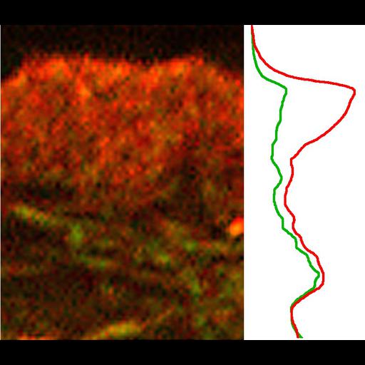
Localization of cross-linking proteins in fibroblast cytoskeleton. Fluorescence microscopy and corresponding intensity profiles of Xenopus fibroblast lamellipodia double stained with TRITC-phalloidin (red) and α-actinin antibodies (green). The protein/actin ratio at the leading edge of the lamellipodium is low for α-actinin compared with internal actin structures. Immuno-EM of the cell edge (CIL 34927) or interior (CIL 34930) of cytochalsin D-treated Xenopus fibroblasts stained with α-actinin ( primary antibody and 18-nm gold-conjugated secondary antibody) reveals α-actinin at filament crossovers in the cell interior. Image corresponds to Figure 5c and c' from J Cell Biol. 1999 May 31;145(5):1009-26.
Procedures for detergent extraction, immunostaining, S1 decoration, light, and EM were described previously (Svitkina et al., 1995, 1996, 1997;Verkhovsky et al., 1995; Svitkina and Borisy, 1998).
| Spatial Axis | Image Size | Pixel Size |
|---|---|---|
| X | 784px | 0.098µm |
| Y | 678px | 0.098µm |