Licensing
Description
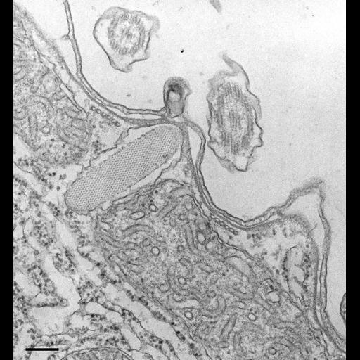
Mucocysts have a herringbone pattern of subunits and are surrounded by a membrane that fuses with the plasma membrane. When the mucocyst content is extruded the content expands (see Hausmann, Protistologica 8:401-412, 1972 for details. TEM taken on 8/12/67 by R. Allen with Philips 200 operating at 60kV. Neg. 19,200X. Bar = 0.2µm. The negative was printed to paper and the image was scanned to Photoshop. This digitized image is available for qualitative analysis. An unprocessed, high resolution version of this image (CIL:34731) is in the library and available for quantitative analysis. Additional information available at (http://www5.pbrc.hawaii.edu/allen/).
Technical Details
Standard glutaraldehyde fixation followed by osmium tetroxide, dehydrated in alcohol and embedded in an epoxy resin. Microtome sections prepared at approximately 75nm thickness. Additional information available at (http://www5.pbrc.hawaii.edu/allen/).
Biological Sources
- NCBI Organism Classification
- Tetrahymena pyriformis
- (Protozoans)
- Cell Type
- cell by organism
- eukaryotic cell
- Eukaryotic Protist
- Ciliated Protist
- Cellular Component
- cell cortex
- cell cortex part
- extrusome
Biological Context
- Biological Process
- cortical cytoskeleton organization
Attribution
- Name
- Richard Allen (University of Hawaii)
- Link
- http://www5.pbrc.hawaii.edu/allen/
Grouping
This image is part of a group.Imaging
- Image Type
- transmission electron microscopy (TEM)
- illumination by electrons
- Imaging Mode
- detection of electrons
- film
- Parameters Imaged
- electron density
- Source of Contrast
- intrinsic mass distribution
- stain with broad specificity
- Visualization Methods
- stain with broad specificity
- uranyl salt
- gold
- Processing History
- recorded image
- film
- Print from negative scanned for Photoshop.
- Data Qualifiers
- processed data
Sample Preparation
- Methods
- glutaraldehyde fixed tissue
- tissue in vitreous ice embedment
- microtome-sectioned tissue
- monoclonal antibody
- Relation To Intact Cell
- microtome-sectioned tissue
Dimensions
| Spatial Axis | Image Size | Pixel Size |
|---|---|---|
| X | 1647px | —— |
| Y | 1784px | —— |