Alternate header for print version


Licensing
This image is copyright protected. Any public or private use of this image is subject to prevailing copyright laws. Please contact the content provider of this image for permission requests.
tweet  
*CIL – Cell Image Library accession number. Please use this to reference an image.

CIL:34539*  Cite 
Description

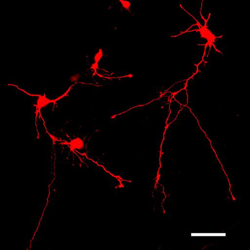
Cortical mouse neurons at 7 days in vitro 7 (DIV7) are well polarized with a single, long, branched axon and shorter dendrites arising from the cell body. The cortical neurons were cultured from E17 mouse embryo and tranfected with DsRed expression plasmid (BD Biosciences). Scale bar, 50 µm.

Technical Details

Cortical neurons were isolated and cultured from E17 embryo using a standard protocol as described in Lehnardt et al.,Proc Natl Acad Sci U S A, 100 (14): 8514-9. Neurons were transfected with a plasmid encoding DsRed (BD Biosciences) at day-in-vitro (DIV) 3. Cells were fixed with 4% paraformaldehyde at DIV5, and images were digitally acquired using a fluorescence microscope (Nikon Eclipse 660) equipped with Spot cooled CCD camera (Diagnostic Instruments).

Biological Sources
NCBI Organism Classification
Mus musculus
Cell Type
CNS neuron (sensu Vertebrata)
Cellular Component
axon
dendrite
Biological Context
Biological Process
neuron development
establishment of cell polarity
Attribution
Names
Yinghua Ma
Timothy Vartanian
Citation
Digital Object Identifier (DOI)
doi:10.7295/W9CIL34539
Archival Resource Key (ARK)
ark:/b7295/w9cil34539
Imaging
Image Type
recorded image
Image Mode
fluorescence microscopy
Parameters Imaged
fluorescence emission
Source of Contrast
distribution of a specific protein
Visualization Methods
DsRed
Data Qualifiers
processed data
suitable for spatial measurements
Sample Preparation
Methods
formaldehyde fixed tissue
Relation To Intact Cell
dispersed cells in vitro
Dimensions
Spatial Axis Image Size Pixel Size
X 3740px 0.089µm
Y 4000px 0.089µm