Licensing

Pd
Public Domain: This image is in the public domain and thus free of any copyright restrictions. However, as is the norm in scientific publishing and as a matter of courtesy, any user should credit the content provider for any public or private use of this image whenever possible. Learn more
Comments: Powered by Pnyxe

Description

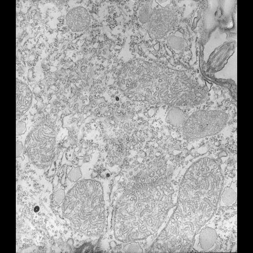
High resolution image of the cytoplasm of Tetrahymena. Mitochondria have tubular cristae, infoldings of their inner membranes. Peroxisomes, with a few tubular inclusions, mucocysts, and a planar view of rough ER, to which many polysomal ribosome arrays are attached, fill the cytosol. TEM taken on 2/11/71 by R. Allen with Hitachi HU11A operating at 75kV. Neg. 19,500X. The raw film was scanned with an Epson Perfection V750 Pro. This image is best used for quantitative analysis.

Technical Details

Standard glutaraldehyde fixation followed by osmium tetroxide, dehydrated in alcohol and embedded in an epoxy resin. Microtome sections prepared at approximately 75nm thickness. Additional information available at (http://www5.pbrc.hawaii.edu/allen/).

Biological Sources

NCBI Organism Classification
Tetrahymena pyriformis
(Protozoans)
Cell Type
cell by organism
eukaryotic cell
Eukaryotic Protist
Ciliated Protist
Cellular Component
cytoplasm
mitochondrion
mitochondrial inner membrane
peroxisome
rough endoplasmic reticulum
polysome

Biological Context

Biological Process
cytoplasm organization

Attribution

Name
Richard Allen (University of Hawaii)
Link
http://www5.pbrc.hawaii.edu/allen/

Grouping

This image is part of a group.
Data Qualifiers
raw, unprocessed data
suitable for spatial measurements

Dimensions

Spatial Axis Image Size Pixel Size
X 4687px 0.77nm
Y 5356px 0.77nm
*CIL – Cell Image Library accession number. Please use this to reference an image.