Licensing

Pd
Public Domain: This image is in the public domain and thus free of any copyright restrictions. However, as is the norm in scientific publishing and as a matter of courtesy, any user should credit the content provider for any public or private use of this image whenever possible. Learn more
Comments: Powered by Pnyxe

Description

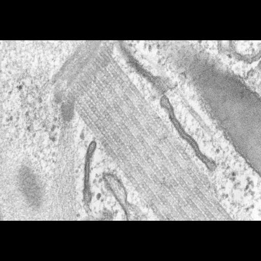
A high resolution image of a bundle of microtubules supporting the oral apparatus of Coleps. This micrograph shows the detail of microtubular cross-bridging within one of these rods. Microfibrillar bridges are located at 22nm intervals within the hexagonally packed microtubules. Standard glutaraldehyde fixation followed by osmium tetroxide, dehydrated in alcohol and embedded in an epoxy resin. Microtome sections prepared at approximately 75nm thickness. TEM taken on 5/29/69 by R. Allen with Philips 300 operating at 60kV. Neg. 63,750X. The raw film was scanned with a Nikon Coolscan 9000ED. This high resolution image is suitable for quantitative analysis. Additional information available at (http://www5.pbrc.hawaii.edu/allen/).

Biological Sources

NCBI Organism Classification
Coleps hirtus
Cell Type
eukaryotic cell
Eukaryotic Protist
Ciliated Protist
Cell Line
Cambridge Culture Collection, Cambridge, England, catalog # LB1656/1
Cellular Component
microtubule associated complex
microtubular crossbridging

Attribution

Name
Richard Allen
Link
http://www5.pbrc.hawaii.edu/allen/

Grouping

This image is part of a group.
Data Qualifiers
raw, unprocessed data
suitable for spatial measurements

Dimensions

Spatial Axis Image Size Pixel Size
X 4039px 0.3nm
Y 2798px 0.3nm
*CIL – Cell Image Library accession number. Please use this to reference an image.