Licensing
Description
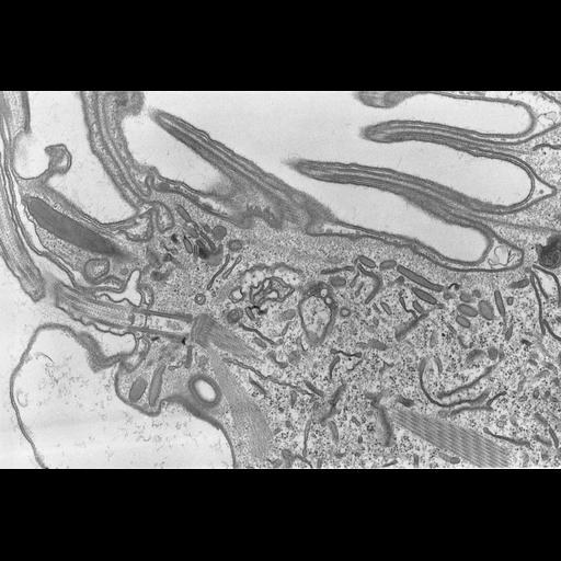
A high resolution image of a longitudinal section of an oral basal body and cilium with the attachment of a bundle of microtubular filaments and a rod that covers the proximal end of the basal body. A layer of filaments underlies the corrugated lips and lamellae extend from these lips. The oblique section through the bundle of microtubules exposes the periodic appearance of the bundle. Parasomal sacs, discoidal vesicles, mucocysts, early endosomes, and toxicysts are seen in this cortical section. Standard glutaraldehyde fixation followed by osmium tetroxide, dehydrated in alcohol and embedded in an epoxy resin. Microtome sections prepared at approximately 75nm thickness. TEM taken on 5/28/69 by R. Allen with Philips 300 operating at 60kV. Neg. 14,800X. The raw film was scanned with a Nikon Coolscan 9000ED. This high resolution image is suitable for quantitative analysis Additional information available at (http://www5.pbrc.hawaii.edu/allen/).
Biological Sources
- NCBI Organism Classification
- Coleps hirtus
- Cell Type
- eukaryotic cell
- Eukaryotic Protist
- Ciliated Protist
- Cell Line
- Cambridge Culture Collection, Cambridge, England, catalog # LB1656/1
- Cellular Component
- cilium axoneme
- axonemal microtubule
- axonemal dynein complex
- plasma membrane
- microtubule basal body
- cytoskeleton
- microtubule cytoskeleton
- actin cytoskeleton
Biological Context
- Biological Process
- ciliary or flagellar motility
- microtubule cytoskeleton organization
- microfibril organization
- aid to ingestion
- Molecular Function
- structural molecule activity
- structural constituent of cytoskeleton
- motility
Attribution
- Name
- Richard Allen
- Link
- http://www5.pbrc.hawaii.edu/allen/
Grouping
This image is part of a group.Imaging
- Image Type
- transmission electron microscopy (TEM)
- illumination by electrons
- Imaging Mode
- detection of electrons
- film
- Parameters Imaged
- electron density
- Source of Contrast
- stain with broad specificity
- Visualization Methods
- stain with broad specificity
- osmium tetroxide
- uranyl salt
- lead salt
- Processing History
- recorded image
- film
- unprocessed raw data
- Data Qualifiers
- raw, unprocessed data
- suitable for spatial measurements
Sample Preparation
- Methods
- glutaraldehyde fixed tissue
- osmium tetroxide fixed tissue
- tissue in epoxy resin embedment
- Relation To Intact Cell
- microtome-sectioned tissue
Dimensions
| Spatial Axis | Image Size | Pixel Size |
|---|---|---|
| X | 5582px | 1.0nm |
| Y | 3764px | 1.0nm |