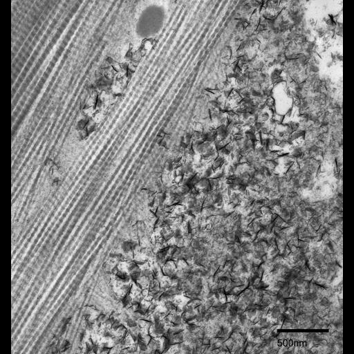
Collagen fibrils and mineralization in the external layer of Fundulus heteroclitus scale. The external layer is the second of three layers closest to the dermis of an elasmoid. It is composed of thin collagen fibrils about 30nm in diameter, which are organized in a loose meshwork. This layer is the first layer to be formed and mineralized. This mineralization is visible in the extracellular material surrounding the collagen fibrils where mineral deposits in the form of needle-like crystals identified to most likely be hydroxyapatite can be found.
Fundulus heteroclitus scales were chemically fixed with 2.5% glutaraldehyde, 2% formaldehyde in 0.1M cacodylate buffer (pH 7.3), then post-fixed in 4% osmium tetroxide and stained en bloc in 1% uranyl acetate. The scales were then dehydrated in a graded series of ethanol and infiltrated with Spurr’s resin. Thin sections of 70 nm were trimmed using a diamond knife and post-stained in uranyl acetate and lead citrate. This micrograph was imaged using a Phillips CM 100 transmission electron microscope at an accelerating voltage of 80 kV.
| Spatial Axis | Image Size | Pixel Size |
|---|---|---|
| X | 1689px | 1.87nm |
| Y | 1798px | 1.87nm |