Alternate header for print version


Licensing
Public Domain: This image is in the public domain and thus free of any copyright restrictions. However, as is the norm in scientific publishing and as a matter of courtesy, any user should credit the content provider for any public or private use of this image whenever possible. Learn more
tweet  
*CIL – Cell Image Library accession number. Please use this to reference an image.

CIL:27225*  Cite 
Description

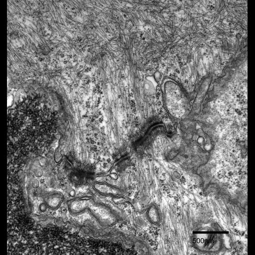
Cell junctions formed between adjacent epidermal cells in Fundulus heteroclitus scales. Desmosomes line the membranes of two epidermal cells. A vast network of intermediate filaments and microfilaments are present throughout the cytoplasm and connect to the desmosomes plates. Microtubules are also visible in the cells.

Technical Details

Fundulus heteroclitus scales were chemically fixed with 2.5% glutaraldehyde, 2% formaldehyde in 0.1M cacodylate buffer (pH 7.3), then post-fixed in 4% osmium tetroxide and stained en bloc in 1% uranyl acetate. The scales were then dehydrated in a graded series of ethanol and infiltrated with Spurr’s resin. Thin sections of 70 nm were trimmed using a diamond knife and post-stained in uranyl acetate and lead citrate. This micrograph was imaged using a Phillips CM 100 transmission electron microscope at an accelerating voltage of 80 kV.

Biological Sources
NCBI Organism Classification
Fundulus heteroclitus
Cell Type
epidermal cell
Cellular Component
cell-cell junction
desmosome
intermediate filament
microfilament
microtubule
Biological Context
Biological Process
cytoskeleton organization
cell-cell junction organization
Attribution
Names
Wai Yan Lam
Marian Rice
Citation
Digital Object Identifier (DOI)
doi:10.7295/W9CIL27225
Archival Resource Key (ARK)
ark:/b7295/w9cil27225
Grouping This image is part of a group.
Imaging
Image Type
recorded image
Image Mode
transmission electron microscopy (TEM)
Parameters Imaged
electron density
Source of Contrast
differences in adsorption or binding of stain
Visualization Methods
uranyl salt
lead salt
Processing History
unprocessed raw data
Dimensions
Spatial Axis Image Size Pixel Size
X 1706px 1.92nm
Y 1808px 1.92nm