Alternate header for print version


Licensing
Attribution Share Alike:This image is licensed under a Creative Commons Attribution, Share Alike License. View License Deed | View Legal Code
tweet  
*CIL – Cell Image Library accession number. Please use this to reference an image.

CIL:26589*  Cite 
Description

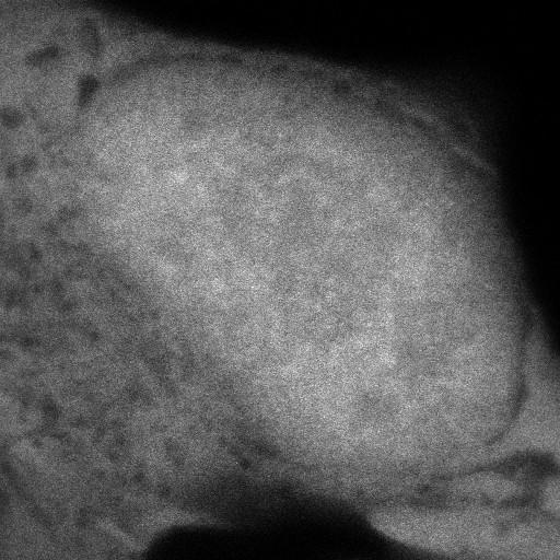
HeLa cells stably expressing BAC-encoded GFP and imaged witb a PicoQuant MicroTime 200 confocal microscope using an Olympus 60x 1.2NA water immersion objective lens. The GFP is distributed throughout the nucleoplasm. From Fig 2A in Huranova et al. 2010 J Cell Biol 191:75-86.

Biological Sources
NCBI Organism Classification
Homo sapiens
Cell Type
cervical epithelium
Cell Line
HeLa
Cellular Component
pre-mRNA
snRNP
spliceosomal complex
Biological Context
Biological Process
RNA splicing
RNA metabolic process
Attribution
Names
M. Huranova
I. Ivani
A. Benda
I. Poser
Y. Brody
M. Hof
Y. Shav-Tal
K.M. Neugebauer
D. Stanek
Published
M Huranova et al. 2010 J Cell Biol 191:75-86
Citation
Digital Object Identifier (DOI)
doi:10.7295/W9CIL26589
Archival Resource Key (ARK)
ark:/b7295/w9cil26589
Grouping This image is part of a group.
Imaging
Image Type
recorded image
Image Mode
confocal microscopy
Parameters Imaged
fluorescence emission
Source of Contrast
distribution of a specific protein
Visualization Methods
EGFP
Processing History
line averaging
Data Qualifiers
processed data
Sample Preparation
Methods
living tissue
Relation To Intact Cell
whole mounted tissue
Dimensions
Spatial Axis Image Size Pixel Size
X 512px ——
Y 512px ——