Licensing
Description
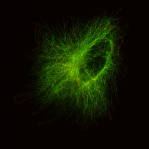
RCC-4 cell expressing wild-type Von Hippel-Lindau protein (pVHL30) stained with anti-alpha-tubulin (green) and anti-GTP-tubulin (red) antibodies. GTP-tubulin localizes to the microtubule (MT) tip and remnants of GTP-tubulin are found along the entire MT lattice. When pVHL30 is expressed in RCC-4 cells, a higher frequency of GTP caps and GTP remnants is observed relative to RCC-4 control (VHL negative). RCC-4 cells were permeabilized in PEM buffer with glycerol and 0.1% Triton-X100, then incubated with primary antibody hMB11 followed by incubation with secondary antibody. Cells were then fixed in cold methanol for 5 min and stained with the polyclonal anti-tubulin antibody followed by secondary antibody. Stacks of images were acquired with an IX70 Delta Vision Spectris microscope using a 60X NA1.4 DIC oil Plan-Apochromat objective. This stack corresponds to J Cell Biol. 2010. 190: 991-1003--Fig 5F (right 2 panels) and the control (left 2 panels in Fig 5F) is CIL# 26572.
Biological Sources
- NCBI Organism Classification
- Homo sapiens
- (human)
- Cell Type
- renal cell carcinoma
- epithelial cell
- Cell Line
- RCC4
- Cellular Component
- microtubule
- cytoskeleton
- protein complex
Biological Context
- Biological Process
- protein polymerization
- Molecular Function
- GTPase activity
- structural molecule activity
- protein binding
Attribution
- Names
- Claudio R. Thoma
- Alexandre Matov
- Katrin L. Gutbrodt
- Christian R. Hoerner
- Zlatko Smole
- Wilhelm Krek
- Gaudenz Danuser
- Published
- J Cell Biol. 2010. 190: 991-1003.
- Link
- http://jcb-dataviewer.rupress.org/jcb/...
- Pubmed
- 20855504
Grouping
This image is part of a group.Imaging
- Image Type
- recorded image
- Imaging Mode
- widefield illumination
- Parameters Imaged
- fluorescence emission
- Source of Contrast
- distribution of a specific protein
- Visualization Methods
- primary antibody plus labeled secondary antibody
- Processing History
- unprocessed raw data
- Data Qualifiers
- raw, unprocessed data
- suitable for spatial measurements
Sample Preparation
- Methods
- methanol fixed tissue
- detergent permeabilized
- Relation To Intact Cell
- dispersed cells in vitro
Dimensions
| Spatial Axis | Image Size | Pixel Size |
|---|---|---|
| X | 1024px | .1103μm |
| Y | 1024px | .1103μm |
| Z | 8 | .2μm |