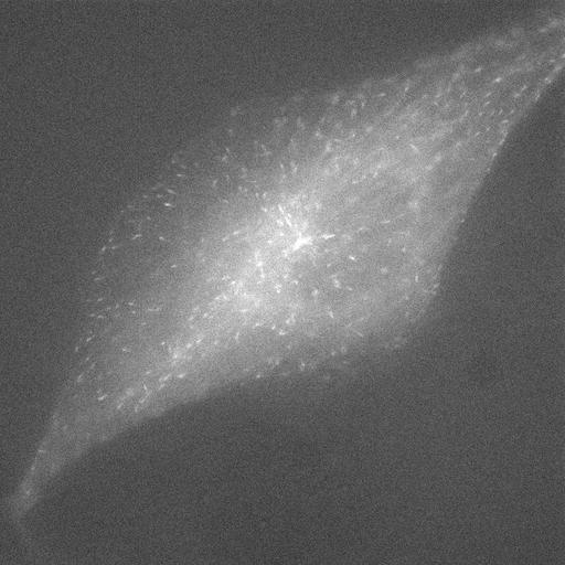
Video shows the dynamics of microtubules (MTs) using a GFP fusion construct of EB3 (end binding protein 3) that transiently binds to growing MT plus ends, generating a punctate pattern of EB3-GFP comets throughout the cell. This video compares the number of EB3-GFP comets detected after nocodazole perturbation in an RCC-4 cell with active VHL. Compare with control video (RCC-4 alone) in CIL # 32024; there is an increased number of growing MTs when VHL is active. RCC-4 cells were infected with a retroviral vector to express EB3-GFP. Cells on round coverslips were transferred to a homemade holding device, and 600 µl of the appropriate medium, containing 40 nM nocodazole was added. The video was acquired with a microscope (IX70 Delta Vision Spectris; Olympus), temperature-controlled at 37C, using a 60× NA 1.4 differential interference contrast (DIC) oil Plan-Apochromat objective, ex 470 em 520, and a camera (CoolSNAP HQ; Roper Industries) with an exposure time of 100 ms and a frame rate of 0.5 s. Acquisition software used was SoftWoRx version 3.3.4 (Applied Precision). This is the raw image file for the right half of Video 5 in J Cell Biol. 2010. 190: 991-1003.
| Spatial Axis | Image Size | Pixel Size |
|---|---|---|
| X | 768px | 0.1103µm |
| Y | 768px | 0.1103µm |
| Time | 0.5 seconds | 80 |
|---|