Alternate header for print version


Licensing
Attribution Non-Commercial Share Alike:This image is licensed under a Creative Commons Attribution, Non-Commercial Share Alike License. View License Deed | View Legal Code
tweet  
*CIL – Cell Image Library accession number. Please use this to reference an image.

CIL:26545*  Cite 
Description

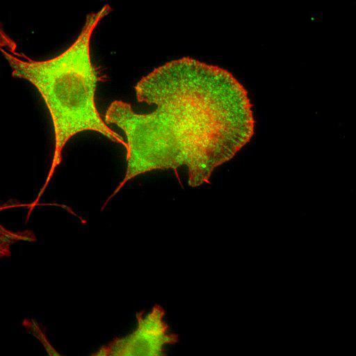
To study the molecular mechanism by which nonmuscle myosin II (MII) regulates protrusion and adhesion dynamics in migrating cells, NIH3T3 cells were treated with scrambled siRNA (Scr) for 2 days, treated with DMSO for 30 min., and stained for βPIX (green) and actin (red). These findings help elucidate a functional link between MII and Rac1/Cdc42 GTPases, which may regulate protrusion/adhesion dynamics in migrating cells. This image is the original data file from Fig. 6B, “Requirement for βPIX in MII-regulated cell protrusion and adhesion.” J. Cell Biol. 2010. Vol. 190(4):663–674.

Technical Details

Cells were cultured in DME (Invitrogen) supplemented with 10% fetal bovine serum and 100 U/ml penicillin/streptomycin (Invitrogen) at 37°C in a humidified 5% CO2 incubator. Scrambled siRNA transfected NIH3T3 cells were treated with DMSO for 30 min, and co-stained for betaPIX (green) and actin (phalloidin, red). Images were captured by Olympus IX81-ZDC inverted microscope with UPlanFL N 40X objective.

Biological Sources
NCBI Organism Classification
Mus musculus
Cell Type
fibroblast
Cell Line
NIH/3T3
Cellular Component
lamellipodium
myosin II complex
actin filament
Biological Context
Biological Process
regulation of cell migration
Attribution
Names
Chan-Soo Lee
Chang-Ki Choi
Eun-Young Shin
Martin Alexander Schwartz
Eung-Gook Kim
Published
J. Cell Biol. 2010. Vol. 190(4):663–674
Pubmed
PMID:20713598
Link
http://jcb-dataviewer.rupress.org/jcb/browse/...
Citation
Digital Object Identifier (DOI)
doi:10.7295/W9CIL26545
Archival Resource Key (ARK)
ark:/b7295/w9cil26545
Grouping This image is part of a group.
Imaging
Image Type
recorded image
Image Mode
fluorescence microscopy
Parameters Imaged
fluorescence emission
Source of Contrast
distribution of epitope
Visualization Methods
Alexa Fluor 488
Tetramethyl rhodamine (TRITC)
phalloidin
Data Qualifiers
raw, unprocessed data
Sample Preparation
Methods
formaldehyde fixed tissue
detergent permeabilized
Relation To Intact Cell
dispersed cells in vitro
Dimensions
Spatial Axis Image Size Pixel Size
X 512px ——
Y 512px ——