Licensing
Description
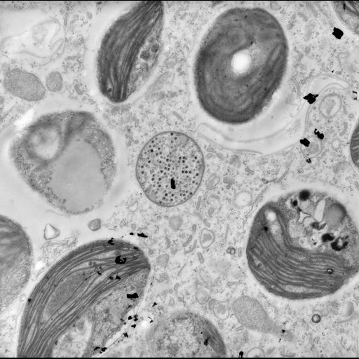
Cytoplasm of a green Trichodina that lives on the surface of a freshwater snail collected in central Illinois. In this view the abundant endosymbiotic zoochlorellae and a single endosymbiotic bacterium are evident. The bacterial endosymbiont is filled with virion particles. In related images the particles can also be seen loose in the cytoplasm of Trichodina. See the serial section CIL:25687). TEM taken by G. Antipa in 1968 on a Hitachi HU11A operating at 75kV. The negative magnification is 11,500X. The raw film was scanned with an Epson Perfection V750 Pro. This image is best used for quantitative analysis. Standard glutaraldehyde fixation followed by osmium tetroxide, dehydrated in alcohol and embedded in an epoxy resin. Microtome sections prepared at approximately 65nm thickness.
Biological Sources
- NCBI Organism Classification
- Trichodina
- Chlorella
- Cell Type
- cell by organism
- eukaryotic cell
- Eukaryotic Protist
- Ciliated Protist
- Cellular Component
- symbiont-containing vacuole
- zoochlorellae
- cytoplasm
- symbiont-containing vacuole
- bacterial symbiont
- virion
- chloroplast thylakoid
- chloroplast thylakoid membrane
Biological Context
- Biological Process
- response to symbiont
- oral apparatus organization
- cytoplasm organization
- eukaryotic photosynthetic symbiont
- bacterial endosymbiont
- viral particles within a bacterial symbiont
- chloroplast organization
Attribution
- Name
- Gregory Antipa (San Francisco State University)
- Link
- http://online.sfsu.edu/~antipa/
Grouping
This image is part of a group.Imaging
- Image Type
- transmission electron microscopy (TEM)
- illumination by electrons
- Imaging Mode
- detection of electrons
- film
- Parameters Imaged
- electron density
- Source of Contrast
- stain with broad specificity
- Visualization Methods
- stain with broad specificity
- osmium tetroxide
- uranyl salt
- lead salt
- Processing History
- recorded image
- film
- unprocessed raw data
- Data Qualifiers
- raw, unprocessed data
- suitable for spatial measurements
Sample Preparation
- Methods
- glutaraldehyde fixed tissue
- osmium tetroxide fixed tissue
- tissue in epoxy resin embedment
- microtome-sectioned tissue
- Relation To Intact Cell
- microtome-sectioned tissue
Dimensions
| Spatial Axis | Image Size | Pixel Size |
|---|---|---|
| X | 5193px | 1.3nm |
| Y | 5164px | 1.3nm |