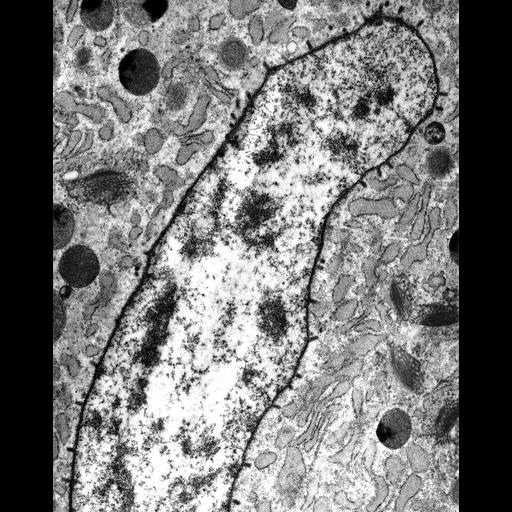
Electron micrograph of Maize mosaic virus (MMV, Rhabdoviridae) in the accessory salivary gland of the insect vector Peregrinus maidis (planthopper, Hemiptera, Delphacidae). MMV virions are bullet-shaped or bacilliform, 186-213 nm long and ca. 61 nm in diameter. MMV buds through nuclear membranes and accumulates in perinuclear space in cells of the accessory salivary gland. The accompanying image in this image group includes labels and scale bar. Methods: Dissected insect organs were processed for TEM by fixation in glutaraldehyde and osmium tetroxide and embedded in Spurr's medium. Thin sections were stained with uranyl acetate and lead citrate, and examined by a Philips-201 TEM. Associated references: Ammar, E.-D. and S. A. Hogenhout, S.A. (2008). A neurotropic route for Maize mosaic virus (Rhabdoviridae) in its planthopper vector Peregrinus maidis. Virus Research 131: 77-85. Ammar, E.-D. and Nault, L.R. (1985). Assembly and accumulation sites of Maize mosaic virus in its planthopper vector. Intervirology 24: 33-41.
| Spatial Axis | Image Size | Pixel Size |
|---|---|---|
| X | 797px | 0.01µm |
| Y | 1006px | 0.01µm |