Licensing

Attribution_nc_sa
Attribution Non-Commercial Share Alike:This image is licensed under a Creative Commons Attribution, Non-Commercial Share Alike License. View License Deed | View Legal Code
Comments: Powered by Pnyxe

Description

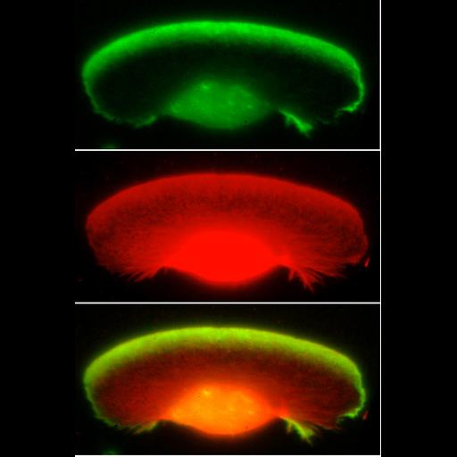
Localization of Arp2/3 complex in the lamellioidia of a Xenopus keratocytes. Staining with p21 antibody (green) and TRIT-phalloidin (red) shows Arp2/3 complex highly enriched in lamellipodia. CIL 24795 shows an immuno EM image of Arp2/3 localization in a Xenopus keratocyte. CIL 24796 and 24797 show corresponding images in fibroblasts. Image corresponds to Figure 3a-c from J Cell Biol. 1999 May 31;145(5):1009-26.

Technical Details

Procedures for detergent extraction, immunostaining, S1 decoration, light, and EM were described previously (Svitkina et al., 1995, 1996, 1997;Verkhovsky et al., 1995; Svitkina and Borisy, 1998).

Biological Sources

NCBI Organism Classification
Xenopus laevis
(African clawed frog)
Cell Type
keratocyte
Cellular Component
lamellipodium
actin cytoskeleton

Biological Context

Biological Process
cellular macromolecule localization

Attribution

Names
Tatyana M. Svitkina
Gary G. Borisy
Published
J Cell Biol. 1999 May 31;145(5):1009-26.
Pubmed
10352018

Grouping

This image is part of a group.

Sample Preparation

Methods
glutaraldehyde fixed tissue
methanol fixed tissue
Relation To Intact Cell
dispersed cells in vitro

Dimensions

Spatial Axis Image Size Pixel Size
X 699px 0.126μm
Y 1044px 0.126μm
*CIL – Cell Image Library accession number. Please use this to reference an image.