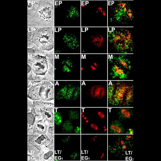
Rat NRK fibroblasts in phase contrast and stained for the nucleolar proteins nucleostemin (green) and fibrillarin (red). Panels at right show merge. Rows show different stages of mitosis, from top, early prophase, late prophase, metaphase, anaphase, telophase, late telophase/early G1.
Data were collected using a Leica DMIRB microscope equipped with a 100x objective (N.A. 1.4) and appropriate filter sets, and images captured using a Quantix 57 CCD camera (Roper Scientific Photometrics). For high resolution spatial mapping, three-dimensional optical stacks (containing 21 consecutive 0.25 micron slices) were captured using a PIFOC microscope focusing drive (Polytec PI). Fluorescence images were dark current subtracted and intensity scaled. See Fig 6 in Politz et al., 2005 Mol Biol Cell 16:3401-3410.
| Spatial Axis | Image Size | Pixel Size |
|---|---|---|
| X | 909px | —— |
| Y | 1380px | —— |