Alternate header for print version


Licensing
Public Domain: This image is in the public domain and thus free of any copyright restrictions. However, as is the norm in scientific publishing and as a matter of courtesy, any user should credit the content provider for any public or private use of this image whenever possible. Learn more
tweet  
*CIL – Cell Image Library accession number. Please use this to reference an image.

CIL:247*  Cite 
Description

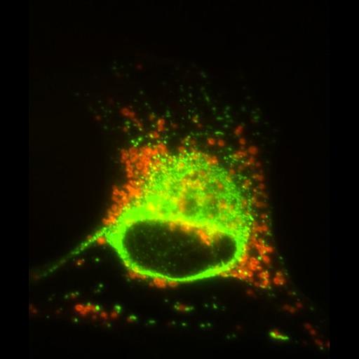
This ghoulish-looking Schwann cell was present in a primary culture of the dorsal root ganglion of mouse that was transfected with a mutant peripherin transgene. The intermediate filament network (vimentin antibody labeling in green) is collapsed around the nucleus 48 hr after transfection, and is correlated with a corresponding peri-nuclear clustering of the mitochondria (labeled with Mitotracker). Mitotracker CMX Ros was added to the culture medium for 30 mins, then rinsed out for 45 minutes in culture before fixing the cells with methanol. Microscope - Zeiss Axioplan I Illumination - Mercury arc lamp Objective - 100X oil/1.3 NA Intermediate lens between obj and camera - 0.63X Camera - Axiocam MRM CCD Filters: 1. Ex 485/20, Em 515-565 BP, dichroic 510 2. Ex 560/40, Em 630/60, dichroic 595

Biological Sources
NCBI Organism Classification
Mus musculus
Cell Type
myelinating Schwann cell
Cellular Component
type III intermediate filament
mitochondrion
Attribution
Names
Parysek, Linda
Citation
Digital Object Identifier (DOI)
doi:10.7295/W9CIL247
Archival Resource Key (ARK)
ark:/b7295/w9cil247
Imaging
Image Type
microscopy
Image Mode
fluorescence microscopy
Parameters Imaged
fluorescence emission
Source of Contrast
local accumulation of fluorescent probe
Visualization Methods
Alexa Fluor 488
CMX rosamine (Mitotracker Red)
primary antibody plus labeled secondary antibody
Processing History
unprocessed raw data
Sample Preparation
Methods
methanol fixed tissue
Relation To Intact Cell
whole mounted tissue
Dimensions
Spatial Axis Image Size Pixel Size
X —— 104nm
Y —— 104nm