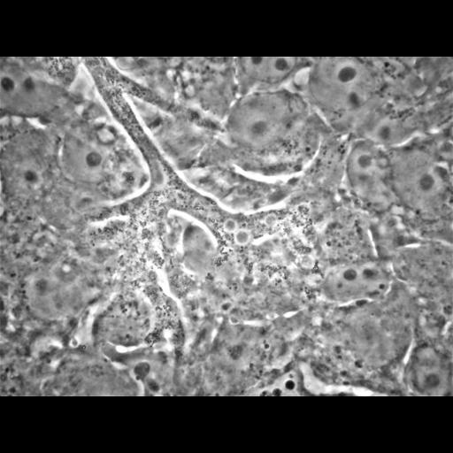
An ameba, Balamuthia mandrillaris, within a culture of feeder cells is extended from its branched pseudopodia at the upper left corner of the picture while its rope-like body curves over and around a nucleus (round circle with three dark rounded spheres, nucleoli) of a monkey kidney cell; additional branches extended both downward and upward into and between other cells. Those branches terminate with more finger-like pseudopodia projections. The density and the movement of the cytoplasmic granules delineate the ameba from its feeder cells that, by comparison, are quiescent with few scattered cytoplasmic granules and a large, rounded nucleus containing several dense nucleoli. The purpose of this movie is to indicate the identity of an ameba amongst other cells in the co-culture. It shows the expanse of the ameba and its simultaneous involvement with several cells. Observations of the amebas under such conditions show their movement and interactions as they feed in a dimension unlike that of the amebas identification in sectioned material obtained from cases of amebic encephalitis. Adapted with permission.
| Spatial Axis | Image Size | Pixel Size |
|---|---|---|
| X | 2272px | —— |
| Y | 1704px | —— |