Alternate header for print version


Licensing
This image is copyright protected. Any public or private use of this image is subject to prevailing copyright laws. Please contact the content provider of this image for permission requests.
tweet  
*CIL – Cell Image Library accession number. Please use this to reference an image.

CIL:24564*  Cite 
Description

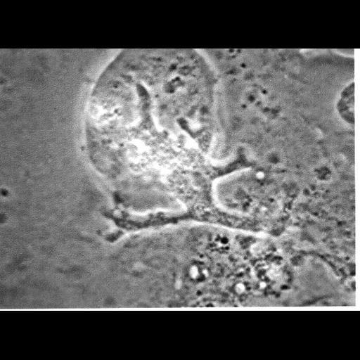
A single frame from the movie (CIL:20154) that shows a large rounded monkey kidney cell being penetrated by a pseudopodium followed by the whole Balamuthia ameba. Detection of the ameba within, rather than on, or beneath the cell is indicated by the change in clarity of the ameba outside and then inside the cell's exterior membrane. The ameba with splayed pseudopodia had begun to enter in apparently a slit in the surface of the cell. The integrity of the cell is maintained and its size does not change appreciably. The portion of the ameba within the cell is less clear than that portion still outside, and as it continues movement within the cell that continues until only a small portion (tail) of the ameba remains outside. Photographed at 1200X. Additional photographs (grouped with this movie) show the movement of the ameba inside of the cell its movement deforms the rounded exterior membrane before the whole ameba emerges some 20 minutes later.

Biological Sources
NCBI Organism Classification
Balamuthia mandrillaris
Cell Type
cell by organism
eukaryotic cell
Eukaryotic Protist
Amoeboid Protist
Cell Line
RP5
Cellular Component
cell
pseudopodium
Attribution
Names
Thelma Dunnebacke
Citation
Digital Object Identifier (DOI)
doi:10.7295/W9CIL24564
Archival Resource Key (ARK)
ark:/b7295/w9cil24564
Grouping This image is part of a group.
Imaging
Image Type
microscopy
in-focus phase contrast
Image Mode
retardance
phase contrast microscopy
Parameters Imaged
boundaries between regions with different refractive index
Source of Contrast
boundaries between regions with different refractive index
Visualization Methods
natural boundaries
Processing History
living tissue
recorded image
charge coupled device (CCD)
Data Qualifiers
raw, unprocessed data
Sample Preparation
Methods
living tissue
Methods
living tissue
whole mounted tissue
Dimensions
Spatial Axis Image Size Pixel Size
X 627px ——
Y 457px ——