Alternate header for print version


Licensing
Attribution Non-Commercial Share Alike:This image is licensed under a Creative Commons Attribution, Non-Commercial Share Alike License. View License Deed | View Legal Code
tweet  
*CIL – Cell Image Library accession number. Please use this to reference an image.

CIL:23047*  Cite 
Description

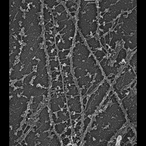
Association of plectin with MTs. Electron microscopy of gelsolin-treated REF-52 cells after immunogold (10 nm) labeling for plectin. Plectin forms bridges between microtubules and intermediate filaments. Electron microscopy of cytoskeletons was performed as described (Svitkina et al., 1995). Briefly, cells on coverslips were lysed as for light microscopy, treated, with recombinant gelsolin NHz-terminal domain, fixed with glutaraldehyde, tannic acid and uranyl acetate, critical point dried, and coated with platinum and carbon. Image corresponds to Fig 5a from J Cell Biol. 1996 Nov;135(4):991-1007.

Biological Sources
NCBI Organism Classification
Rattus
Cell Type
fibroblast
Cell Line
REF-52
Cellular Component
microtubule
intermediate filament
plectin
Biological Context
Biological Process
gelsolin treatment
Attribution
Names
Tatyana M. Svitkina
Alexander B. Verkhovsky,
Gary G. Borisy
Published
J Cell Biol. 1996 Nov;135(4):991-1007
Pubmed
8922382
Citation
Digital Object Identifier (DOI)
doi:10.7295/W9CIL23047
Archival Resource Key (ARK)
ark:/b7295/w9cil23047
Grouping This image is part of a group.
Sample Preparation
Methods
glutaraldehyde fixed tissue
tannic acid
critical_point dried specimen
Relation To Intact Cell
platinum replica
Dimensions
Spatial Axis Image Size Pixel Size
X 2915px ——
Y 3330px ——