Licensing

Pd
Public Domain: This image is in the public domain and thus free of any copyright restrictions. However, as is the norm in scientific publishing and as a matter of courtesy, any user should credit the content provider for any public or private use of this image whenever possible. Learn more
Comments: Powered by Pnyxe

Description

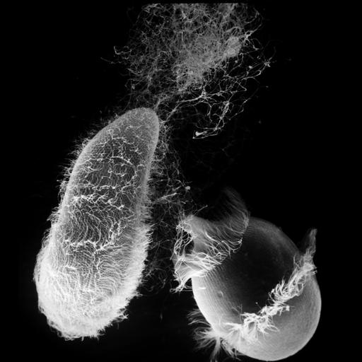
Didinium attacks Paramecium. Also showing many discharged trichocysts and metachronous waves of cilia in the two characteristic ciliary girdles of Didinium nasutum and on Paramecium. A series of capture and ingestion SEMs are grouped with this image. This micrograph was taken in 1968 by G. Antipa on a Cambridge Mark IIA operating at 20kV. The negative magnification is 410X. Further details are available at Wessenberg, H. and Antipa, G. 1970. Capture and ingestion of Paramecium by Didinium nasutum. J. Protozool. 17:250-270.

Biological Sources

NCBI Organism Classification
Didinium nasutum
Paramecium sp.
Cell Type
cell by organism
eukaryotic cell
Eukaryotic Protist
Ciliated Protist
Cellular Component
cell cortex
oral apparatus
trichocyst
cell surface

Biological Context

Biological Process
response to toxin
activation of membrane attack complex

Attribution

Name
Gregory Antipa (San Francisco State University)
Link
http://online.sfsu.edu/~antipa/

Grouping

This image is part of a group.
Data Qualifiers
raw, unprocessed data

Sample Preparation

Methods
chemically fixed tissue
Parducz fixative
lyophilized specimen
Au/Pd metal sputter-coat
Relation To Intact Cell
whole mounted tissue

Dimensions

Spatial Axis Image Size Pixel Size
X 5298px 32nm
Y 5912px 32nm
*CIL – Cell Image Library accession number. Please use this to reference an image.