Alternate header for print version


Licensing
Public Domain: This image is in the public domain and thus free of any copyright restrictions. However, as is the norm in scientific publishing and as a matter of courtesy, any user should credit the content provider for any public or private use of this image whenever possible. Learn more
tweet  
*CIL – Cell Image Library accession number. Please use this to reference an image.

CIL:221*  Cite 
Description

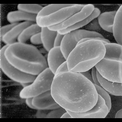
Red blood cells (erythrocytes). This image of human red blood cells obtained by scanning electron microscopy, revealing their characteristic biconcave shape.

Technical Details

The scanning electron microscope scans the specimen with a finely focused electron beam and records the number of electrons emitted at each point in the scan as a grey level in the image, with low levels corresponding to few emitted electrons and high levels corresponding to many emitted. Specimens are usually coated with a thin layer of metal to improve conductivity and increase the efficiency of secondary electron emission.

Biological Sources
NCBI Organism Classification
Homo sapiens
Cell Type
erythrocyte
Biological Context
Biological Process
gas transport
Attribution
Names
Tina Carvalho
Citation
Digital Object Identifier (DOI)
doi:10.7295/W9CIL221
Archival Resource Key (ARK)
ark:/b7295/w9cil221
Imaging
Image Type
recorded image
Image Mode
scanning electron microscopy (SEM)
Parameters Imaged
secondary electron generation
elastic scattering of electrons
Source of Contrast
differences in deposition of metal shadow
Visualization Methods
shadowing and plating
Processing History
unprocessed raw data
Data Qualifiers
processed data
suitable for spatial measurements
Sample Preparation
Methods
critical_point dried specimen
Relation To Intact Cell
whole mounted tissue
Dimensions
Spatial Axis Image Size Pixel Size
X 899px ——
Y 843px ——