Alternate header for print version


Licensing
Public Domain: This image is in the public domain and thus free of any copyright restrictions. However, as is the norm in scientific publishing and as a matter of courtesy, any user should credit the content provider for any public or private use of this image whenever possible. Learn more
tweet  
*CIL – Cell Image Library accession number. Please use this to reference an image.

CIL:214*  Cite 
Description

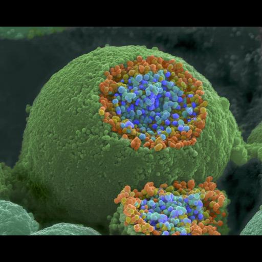
A colorized scanning electron microscope picture of a nerve ending that has been broken open to reveal the synaptic vesicles (orange and blue) beneath the cell membrane.

Biological Sources
Cell Type
neuron
Cellular Component
synaptic vesicle
Biological Context
Biological Process
neuron-neuron synaptic transmission
Attribution
Names
Tina Carvalho
Link
NIGMS "Inside the Cell" booklet
Citation
Digital Object Identifier (DOI)
doi:10.7295/W9CIL214
Archival Resource Key (ARK)
ark:/b7295/w9cil214
Imaging
Image Type
recorded image
Image Mode
scanning electron microscopy (SEM)
Parameters Imaged
secondary_electron imaging
Source of Contrast
differences in deposition of metal shadow
Visualization Methods
shadowing and plating
Processing History
colorized image
Data Qualifiers
processed data
Sample Preparation
Relation To Intact Cell
fractured cell
Dimensions
Spatial Axis Image Size Pixel Size
X 3000px ——
Y 2395px ——