Alternate header for print version


Licensing
This image is copyright protected. Any public or private use of this image is subject to prevailing copyright laws. Please contact the content provider of this image for permission requests.
tweet  
*CIL – Cell Image Library accession number. Please use this to reference an image.

CIL:18042*  Cite 
Description

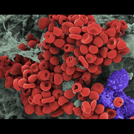
Scanning electron micrograph of a hamster oocyte cumulus complex. Cumulus cells (purple) and matrix (gray) are shown. Small blood clots (red) also often appear in oocyte cumulus complexes. The red blood cells are 6-8 microns in diameter.

Biological Sources
NCBI Organism Classification
Mesocricetus auratus
Cell Type
blood cell
oocyte
cumulus cell
Cellular Component
cell
Biological Context
Biological Process
erythrocyte aggregation
Attribution
Names
Prue Talbot
Citation
Digital Object Identifier (DOI)
doi:10.7295/W9CIL18042
Archival Resource Key (ARK)
ark:/b7295/w9cil18042
Imaging
Image Type
recorded image
Image Mode
scanning electron microscopy (SEM)
Parameters Imaged
elastic scattering of electrons
Source of Contrast
differences in deposition of metal shadow
Visualization Methods
gold
Processing History
colorized with PhotoImpact
Dimensions
Spatial Axis Image Size Pixel Size
X 593px ——
Y 463px ——