Alternate header for print version


Licensing
This image is copyright protected. Any public or private use of this image is subject to prevailing copyright laws. Please contact the content provider of this image for permission requests.
tweet  
*CIL – Cell Image Library accession number. Please use this to reference an image.

CIL:17449*  Cite 
Description

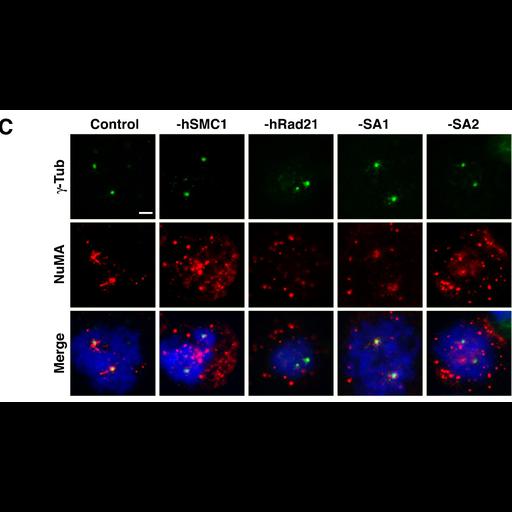
Fluorescence images of control and Si-RNA transfected HeLa cells treated with nocodazole and allowed to recover. Mitotic cells were stained for gamma-tubulin (top row, green)to label centrosomes or NuMA (center row, red). Merges images inclde DAPI staining of DNA (blue). Multiple NuMA signals are not due to centrosome amplification. Cells were observed with an Olympus IX70 microscope and recorded with a Magnaview CCD camera. See Fig 4C in Kong et al., 2009, Mol Biol Cell 20:1289-1301.

Biological Sources
NCBI Organism Classification
Homo sapiens
Cell Type
cervical carcinoma
Cell Line
HeLa
Cellular Component
spindle
kinetochore
nuclear chromosome
Biological Context
Biological Process
mitosis
Attribution
Names
Xiangduo Kong
Alexander Ball
Eiichiro Sonoda
Jie Feng
Shunichi Takeda
Tatsuo Fukagawa
Tim Yen
Kyoko Yokomori
Published
X. Kong et al., 2009 Mol Biol Cell 20:1289-1301
Pubmed
19116315
Link
http://www.molbiolcell.org/cgi/reprint/20/5/1...
Citation
Digital Object Identifier (DOI)
doi:10.7295/W9CIL17449
Archival Resource Key (ARK)
ark:/b7295/w9cil17449
Grouping This image is part of a group.
Imaging
Image Type
charge coupled device (CCD)
Image Mode
fluorescence microscopy
Parameters Imaged
fluorescence emission
Source of Contrast
distribution of a specific protein
differences in adsorption or binding of stain
Visualization Methods
4',6-diamidino-2-phenylindole (DAPI)
Fluorescein (FITC)
Cy5
Processing History
multi-panel montage
Data Qualifiers
processed data
Sample Preparation
Methods
formaldehyde fixed tissue
Relation To Intact Cell
whole mounted tissue
Dimensions
Spatial Axis Image Size Pixel Size
X 1392px ——
Y 796px ——