Alternate header for print version


Licensing
Attribution Non-Commercial Share Alike:This image is licensed under a Creative Commons Attribution, Non-Commercial Share Alike License. View License Deed | View Legal Code
tweet  
*CIL – Cell Image Library accession number. Please use this to reference an image.

CIL:17447*  Cite 
Description

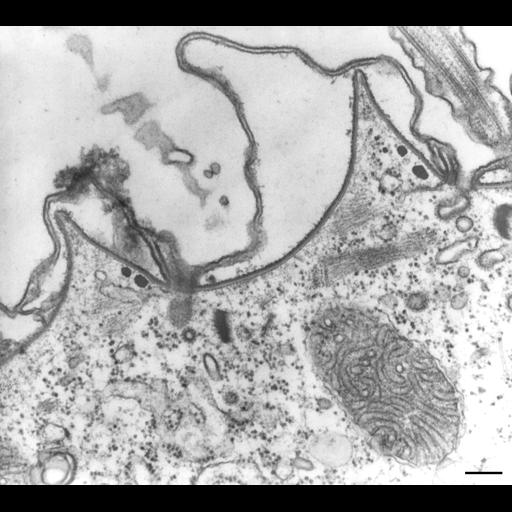
Cell sectioned perpendicular to the surface. The parasomal sac ends internally as a coated pit, coated with clathrin. Where the coated pit has pinched off a clathrin-coated vesicle is formed. When the clathrin is removed an endosome results. Other clathrin-coated vesicles arise from early endosomes. TEM taken on 5/21/70 by R. Allen with Hitachi HU11A operating at 75kV. Neg. 27,750X. Bar = 0.2µm. Adapted with permission from Fig. 1A published in J. Cell Sci. 101:449-461, 1992. The negative was printed to paper and the image was scanned to Photoshop. This digitized image is available for qualitative analysis. An unprocessed, high resolution version of this image (CIL:12612) is in the library and available for quantitative analysis. Standard glutaraldehyde fixation followed by osmium tetroxide, dehydrated in alcohol and embedded in an epoxy resin. Microtome sections prepared at approximately 75nm thickness. Additional information available at (http://www5.pbrc.hawaii.edu/allen/).

Biological Sources
NCBI Organism Classification
Paramecium multimicronucleatum
Cell Type
cell by organism
eukaryotic cell
Eukaryotic Protist
Ciliated Protist
Cellular Component
cell cortex
clathrin coat of endocytic vesicle
cytoplasm
plasma membrane
cortical cytoskeleton
Attribution
Names
Richard Allen (University of Hawaii)
Published
J Cell Sci 101:449-461, 1992
Pubmed
1629255
Citation
Digital Object Identifier (DOI)
doi:10.7295/W9CIL17447
Archival Resource Key (ARK)
ark:/b7295/w9cil17447
Grouping This image is part of a group.
Imaging
Image Type
transmission electron microscopy (TEM)
illumination by electrons
Image Mode
detection of electrons
film
Parameters Imaged
electron density
Source of Contrast
stain with broad specificity
Visualization Methods
stain with broad specificity
osmium tetroxide
uranyl salt
lead salt
Processing History
recorded image
film
Print from negative scanned for Photoshop.
Data Qualifiers
processed data
Dimensions
Spatial Axis Image Size Pixel Size
X 2556px ——
Y 2292px ——