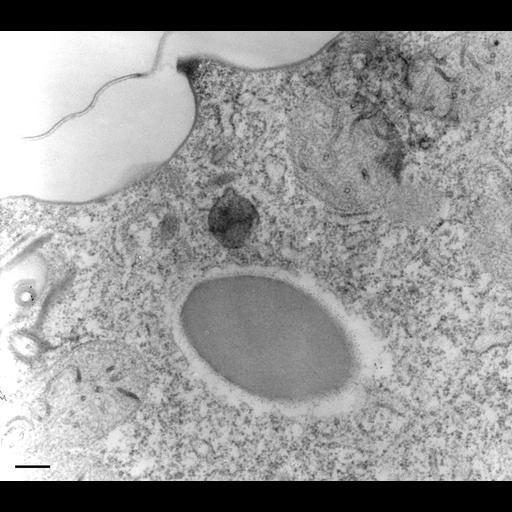
An early endosome in planar view that contains HRP-reaction product. This cell was exposed to HRP for 6 min. and then fixed. This early endosome had two small coated evaginations arising from its rim and also had two large HRP-containing uncoated vesicles docked next to it presumably ready to fuse with it (for this picture and a serial section showing these two vesicles and the second evagination see Fig. 14C in Allen, Schroeder and Fok, J. Cell Sci. 101:449-461, 1992). Mitochondria have an endogenous peroxidase activity. This image was adapted with permission from Fig. 14. TEM taken on 10/20/91 by R. Allen with Zeiss 10A operating at 80kV. Neg. 19,800X. Bar = 0.25µm. The negative was printed to paper and the image was scanned to Photoshop. This digitized image is available for qualitative analysis. An unprocessed, high resolution version of this image (CIL:12617) is in the library and available for quantitative analysis. Standard glutaraldehyde fixation followed by osmium tetroxide, dehydrated in alcohol and embedded in an epoxy resin. Microtome sections prepared at approximately 75nm thickness. Additional information available at (http://www5.pbrc.hawaii.edu/allen/).
| Spatial Axis | Image Size | Pixel Size |
|---|---|---|
| X | 2430px | —— |
| Y | 2139px | —— |