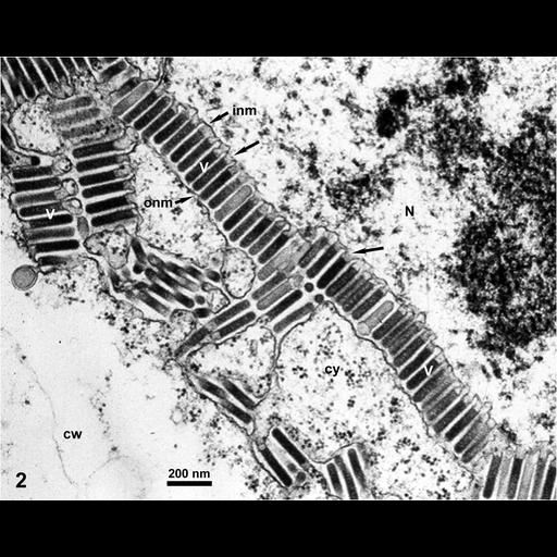
The images in this group show electron micrographs of epidermal and mesophyll cells in thin sections of maize leaves infected with Maize mosaic virus (MMV, Rhabdoviridae). MMV virions are bullet-shaped or bacilliform, 186-213 nm long and ca. 61 nm in diameter. This jpg image is of a mesophyll leaf cell from a maize plant infected with Maize mosaic virus (MMV), and is identical to the unlabeled tif file CIL:12415. MMV virions (v), budding through the inner nuclear membrane (inm), are accumulating between this membrane and the outer nuclear membrane (onm) as well as in intracytoplasmic vesicles, sometimes forming crystalline arrays. Ch=chloroplast; cw=cell wall; cy=cytoplasm; N=nucleus, va=cell vacuole Detailed methods: Pieces of leaves were processed for TEM by fixation in glutaraldehyde and osmium tetroxide, embedded in Spurr's medium. Thin sections were stained with uranyl acetate and lead citrate, and examined by a Philips-201 TEM (non-digital camera). Negatives were scanned later to generate these electronic files. See E.-D. Ammar et al. (2005) J. Phytopathology, 153:129-136.
| Spatial Axis | Image Size | Pixel Size |
|---|---|---|
| X | 2646px | —— |
| Y | 2109px | —— |