Licensing

Attribution_nc_sa
Attribution Non-Commercial Share Alike:This image is licensed under a Creative Commons Attribution, Non-Commercial Share Alike License. View License Deed | View Legal Code
Comments: Powered by Pnyxe

Description

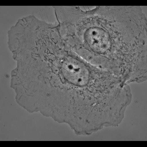
Phase contrast image of control PtK1 cell whose microtubules were visualized by microinjected X-rhodamine tubulin and fluorescence microscopy. This cell corresponds to Figure 1 and Video 1 of J Cell Biol, 161:845-851, 2003. Video 1 is contained within the same image group as this figure.

Biological Sources

NCBI Organism Classification
Potorous tridactylus
(long-nosed potoroo)
Cell Type
epithelial cell
Cell Line
Pt K1 (NBL-3)
Cellular Component
cell
nucleus
lamellipodium
middle lamella

Attribution

Names
Torsten Wittmann
Gary M. Bokoch
Clare M. Waterman-Storer
Published
J Cell Biol, 161:845-851, 2003
Pubmed
12796474

Grouping

This image is part of a group.

Imaging

Image Type
recorded image
Imaging Mode
phase contrast microscopy
Parameters Imaged
elastic scattering of photons
Source of Contrast
differences in amount of elastic light scattering
Visualization Methods
visualization of contiguous regions
Processing History
unprocessed raw data
Data Qualifiers
raw, unprocessed data

Sample Preparation

Methods
living tissue
Relation To Intact Cell
dispersed cells in vitro

Dimensions

Spatial Axis Image Size Pixel Size
X 1030px ——
Y 949px ——
*CIL – Cell Image Library accession number. Please use this to reference an image.