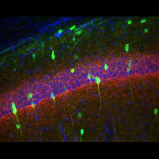
Distribution of interneurons expressing EGFP from the 5HT3 receptor promoter (Tg(Htr3a-EGFP)DH30Gsat, www.gensat.org) colabelled for the CB1 cannabinoid receptor. This image is from the pyramidal cell layer in hippocampal CA1. Multiple EGFP+ bipolar interneurons are present within the pyramidal cell layer and a mesh of EGFP+ fibers and CB1+ axons is present around the pyramidal cell bodies, consistent with basket cell synapses. EGFP+ cells are also present in the stratum oriens and stratum radiatum. CB1 was detected with the L15 antibody (K. Mackie). Section (40 um) was imaged with a 32X, 0.4 NA Achroplan objective on a Zeiss Axiovert microscope. AF568 was excited at 562/40 nm and emission collected at 624/40 nm with Semrock filters and a single band dichroic (530-585 nm reflected, 601-800 nm transmitted). AF488 was excited at 482/35 nm and emission collected at 536/40 nm with Semrock filters and a single band dichroic (446-499 nm reflected, 513-735 nm transmitted). DAPI was excited at 350/50 nm and emission collected at 460/50 nm with a CLP400 dichroic (Chroma). Single images were captured for each channel using a Zeiss Axiocam MR CCD and pseudo-colored using Axiovision. See Davis and Puhl 2011 (Pubmed ID 21305052) for a detailed staining protocol. This image is part of a series characterizing EGFP expression from the 5HT3 promoter throughout the brain.
| Spatial Axis | Image Size | Pixel Size |
|---|---|---|
| X | 1300px | 0.35181496µm |
| Y | 1030px | 0.35181496µm |