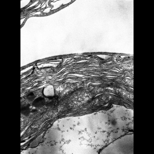
Thin section of Prochloron, a photosynthetic prokaryote showing adpressed thylakoid membranes. This image group includes freeze-fracture images showing the particle sizes and distribution on the thylakoid membranes. See also: Giddings TH, Withers NW, Staehelin LA. 1980 Supramolecular structure of stacked and unstacked regions of the photosynthetic membranes of Prochloron sp., a prokaryote. Proc Natl Acad Sci U S A. 77:352-356.
Prochloron was collected at Coconut Island, Oahu, Hawaii. Green colonies of didemnid , were collected from shallow waters. Green spherical cells, identified as Prochloron, were released from the ascidian Diplosoma virensascidians by gently pressing on the colonies. Material was prepared for thin sectioning as follows: Didemnid colonies containing Prochloron were fixed on site for 2 hr in 1.5% glutaraldehyde in sea water, rinsed, and stored in sea water for 3-4 weeks. Samples were then postfixed in 2% aqueous KMnO4 for 24 hr at room temperature, dehydrated with an acetone series, and embedded in Spurr's resin. Thin sections were stained with aqueous uranyl acetate and lead citrate, and examined in a JEOL 100C TEM at 20,000X magnification. Images were recorded on film and subsequently digitized.
| Spatial Axis | Image Size | Pixel Size |
|---|---|---|
| X | 3385px | 1nm |
| Y | 4608px | 1nm |